OCT provides in vivo detection of bacterial biofilms in critical care patients to reduce the incidence of ventilator‐associated pneumonia.

OCT provides in vivo detection of bacterial biofilms in critical care patients to reduce the incidence of ventilator‐associated pneumonia.

Aged tissues bear the hallmarks of chronic inflammation.
![3D Imaging of Carbon Nanotube Networks [Video]](https://www.advancedsciencenews.com/wp-content/uploads/2019/04/adfm201807901_ASN_image.png)
Quantitative electron tomography provides deeper insight into the 3D topology of carbon nanotube networks.

Researchers study chickpea flour as a potential building block for biodegradable (and edible) packaging materials.
A team of researchers from the University of Paris, France developed a compact high‐speed full‐field optical coherence microscope for high‐resolution in vivo imaging of biological tissues.
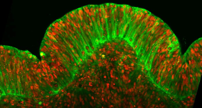
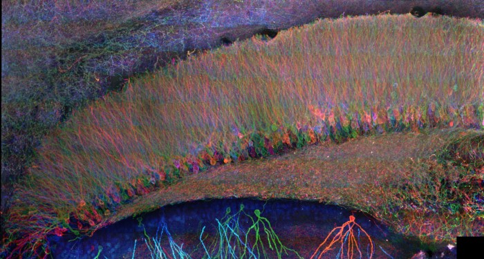
Low cost, on-chip, high-resolution imaging to study cellular dymamics of intact brain organoids.
Step‐by‐step Expansion Microscopy protocols for performing analysis of proteins and RNAs using chemicals and hardware found in a typical biology lab.
![Improving Imaging Techniques in the NIR-II Window [Video]](https://www.advancedsciencenews.com/wp-content/uploads/2018/11/adma201802394_ASN_image.jpg)
The NIR-II window holds great potential in biological imaging.
A visible color change occurs when active nanocomposites are heated and re-cooled, which is attributed to the mobility of the nanoparticles embedded inside the material.

A team of Spanish researchers developed a handheld skin imaging system with joint optical coherence tomography and digital epiluminescence microscopy for point-of-care skin diagnosis.
 New theory suggests gravity is not a fundamental force
 Higgs boson may be driving the Universe’s expansion
 Water-powered gadgets may be on the horizon thanks to new evaporation-based energy device
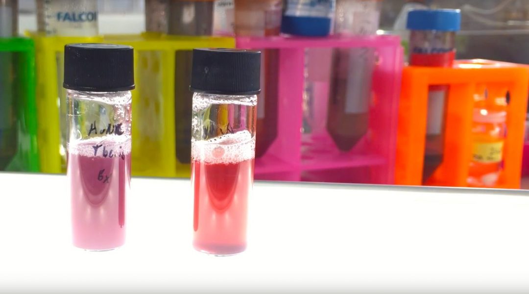
 Scientists discover a new class of antibiotics
 Micron-sized hidden dimensions could solve two of physics’ deepest puzzles
 A new type of dark matter could explain mysterious radiation from the Milky Way’s core