Imaging techniques in the second near-infrared (NIR-II) window are appealing for biological applications because they offer improved image quality compared to conventional methods using light in the visible or first infrared region.
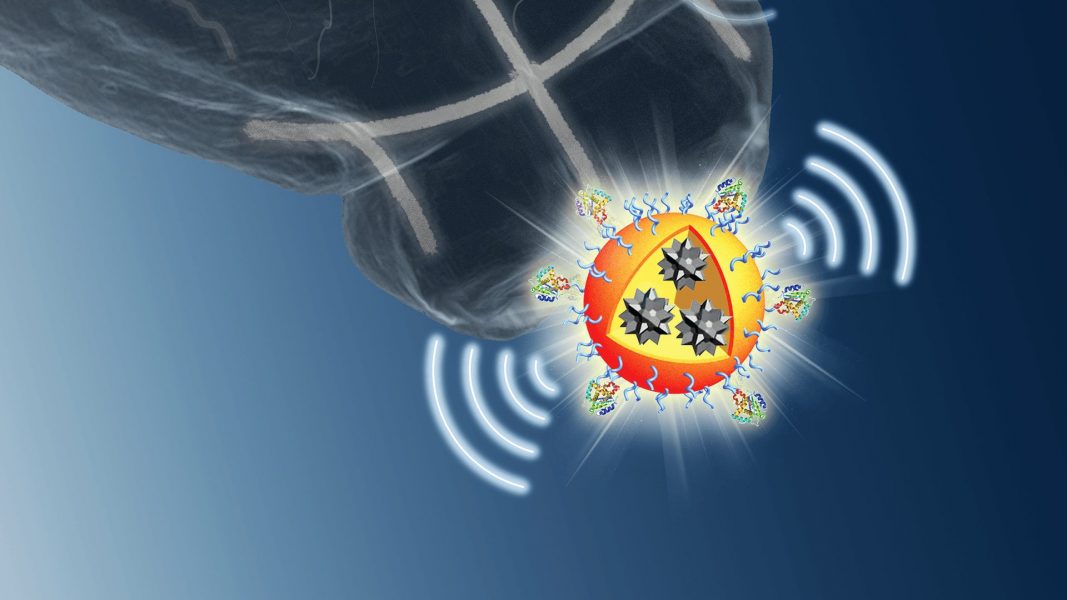
In Advanced Materials, Prof. Bin Liu and co-workers from the National University of Singapore provide an overview of contrast agents for fluorescence and photoacoustic imaging in the NIR-II window.
Imaging in the NIR-II window allows for significantly decreased light scattering by biological tissues, lower background autofluorescence, higher spatiotemporal resolution, and deeper tissue penetration.
Inorganic nanomaterials, including carbon nanotubes and quantum dots, have shown promise for in vivo fluorescence imaging applications. Organic nanomaterials, such as small-molecule fluorophores, especially those with aggregation-induced emission, are emerging NIR-II probes with great potential.
Photoacoustic imaging is a hybrid imaging technique that integrates light excitation with ultrasonic wave detection, which allows for penetration to a depth of several centimetres and significantly enhances the spatial resolution and contrast.
The main challenge we face is not only to design or select effective contrast agents for high-quality imaging, but also to determine their biocompatibility and toxicity effects.
To find out more about NIR-II-emitting contrast agents, please visit the Advanced Materials homepage.


