Full‐field optical coherence microscopy (FF‐OCM), also referred to as full‐field optical coherence tomography (FF‐OCT), is an interferometry imaging technique derived from optical coherence microscopy (OCM), capable of en face tomographic imaging using full‐field illumination with a low‐coherence light source and full‐field detection with an area camera. As there is no depth of field constraint in en face imaging, high numerical aperture (NA) microscope objectives can be used in FF‐OCM to achieve high transverse resolution. High axial resolution can also be obtained in FF‐OCM by using a simple and inexpensive light source such as a halogen lamp or a broadband light‐emitting diode (LED). Cellular‐level imaging with FF‐OCM of biological tissues, ex vivo, has been widely reported. In vivo imaging with FF‐OCM, however, is still challenging.
FF-OCM is subject to so called fringe washout – sample motion that changes the optical phase and then blurs the interferometry signal – as well as FF-OCm havin nog confocal gating mechanism to reject out of-focus-light. Another limitation is the relatively low dynamic range of area cameras, combined with the low radiance of the illumination source. This makes in vivo imaging with FF-OCM difficult unless the sample is carefully immobilized.
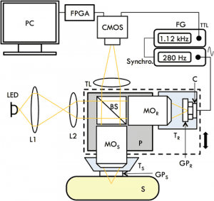
Schematic diagram of the high‐speed compact FF‐OCM prototype
Or you could use a high‐brightness LED and a high‐speed camera.Two French researchers from the Institut d’Optique in Paris developed a FF‐OCM device designed for high‐resolution in vivo imaging that is compact enough to be potentially turned into a handheld device. Full‐field illumination with low‐coherence light was achieved with a high‐brightness broadband light‐emitting diode. High‐speed full‐field detection was established as well, by using part of the image sensor of a high‐dynamic range CMOS camera.
The FF‐OCM device is capable of imaging human skin in vivo without any motion artefact or reconstruction artefacts, at high spatial resolution. Using such a high‐speed imaging device, it is likely that real‐time 3D reconstruction of in vivo samples will soon be possible using FF‐OCM. “Furthermore, the interferometer part of the setup was designed to be as compact and light as possible, while keeping a Linnik interferometer design. This lays the foundations for a handheld device, as the capacity for in vivo imaging is of greater interest if the device is easy to handle” according to scientist Arnaud Dubois.