Albert Einstein once said, “the greatest scientists are artists as well”. This month, our editors gathered some of the most spectacular images from our journals, which capture the beauty and intrigue inherent in science. From evasive microrobots to a dazzling array of crystal polymers, this edition of This month in pictures highlights innovative breakthroughs captured in stunning images.
Yolk–shell structures
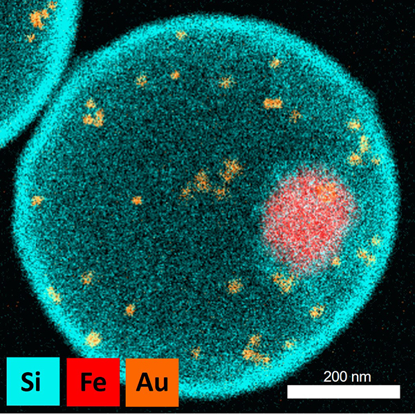
This elemental map of a Fe3O4/AuNS@mSiO2 yolk–shell structure was enabled by energy dispersive X-ray. Jae-Hong Kim, Jae-Hyuk Kim, and co-workers created this image as part of a new strategy for making yolk–shell structured materials. They go on to demonstrate their utility by showing applications in catalysis, photothermal reactions, and sensing.
Shape-shifting magnetic cilia
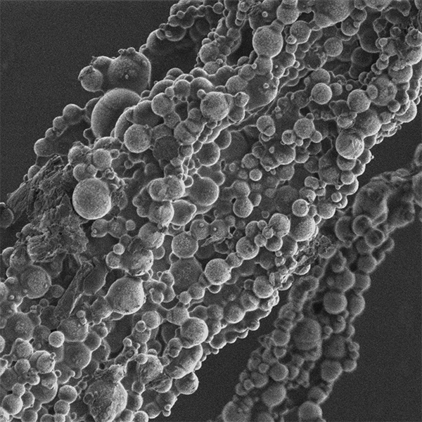
Stimulus‐responsive polymers are attractive for microactuators as they can be easily miniaturized and remotely controlled for untethered operation. This SEM image shows a single magnetic cilium that Joseph Tracy and co-workers prepared through magnetic-assisted self-assembly, and used them to create elastomers with programmable shapes to advance soft materials and robotic devices.
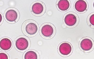
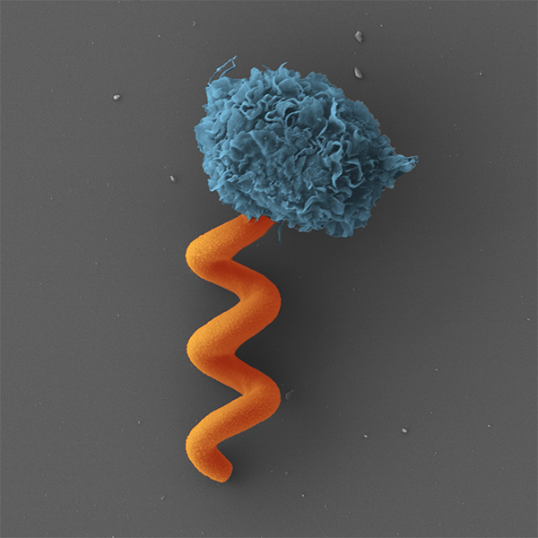
Improving diagnosis
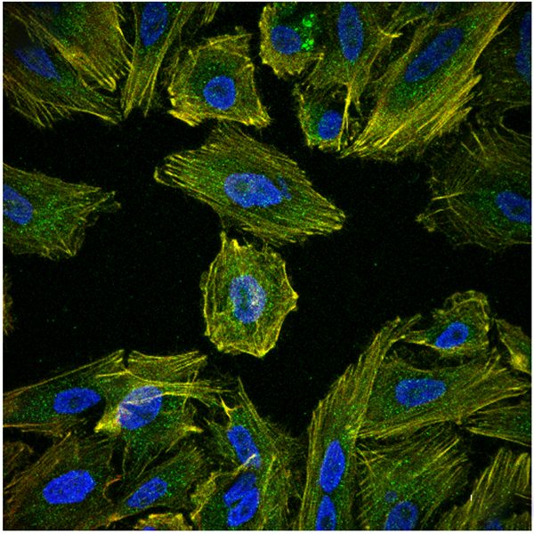
Accurately diagnosing cancer and identifying its stage of development is crucial for saving lives. In a recent study, Anahid Amiri, Florian Hastert, and Christian Dietz show that carcinomas that are likely to spread show a number of distinct characteristics that are detectable through force spectroscopy. Their results could help improve the prognosis in epithelial cancers with metastasis risk.
Light-scattering crystals
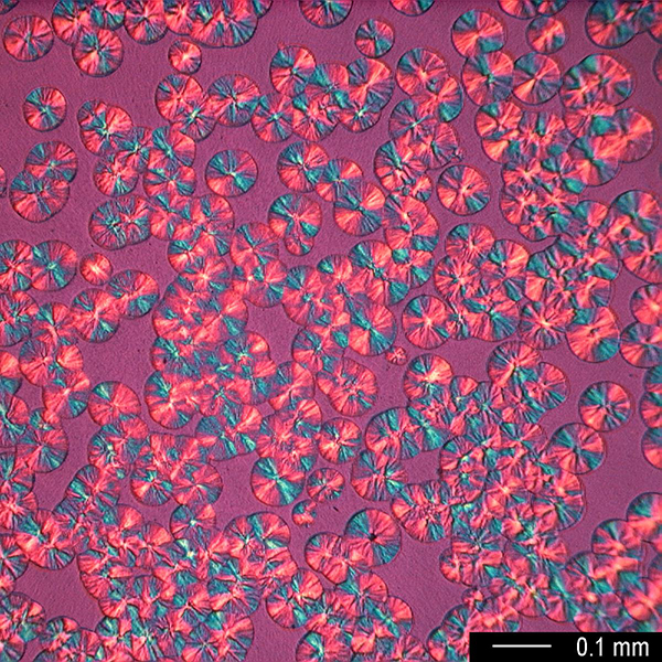
The image above by János Molnfár and co-workers at Budapest University of Technology and Economics shows polycrystaline structures of isotactic polypropylene (iPP) seen through a polarized optical microscope during crystallization at 130 °C. iPP crystals were used as a model for investigating the effects semicrystalline polymers have on light scattering.
Rings of gold
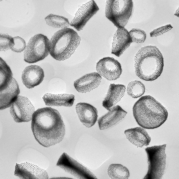
What at first glance look like rings in this TEM image are in fact gold nanoparticles on poly(lactic-co-glycolic acid) (PLGA) spheres. The PLGA spheres are not easily visible, and so the darker gold nanoparticles on the PLGA surface appear to be forming into clusters of rings. Aesthetics aside, the purpose of these PLGA-Au co-assemblies is to enhance photoacoustic imaging and photothermal therapy in treating cancer. This work by Nana Zhao and Fu-Jian Xu and their co-workers demonstrates that the amount of gold nanoparticles on the surface of the PLGA spheres are easily tunable, providing a facile strategy to construct flexible photothermal assemblies with favorable properties for imaging-guided synergistic therapy.
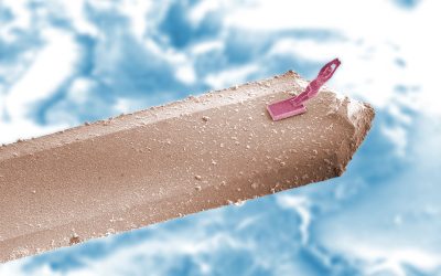
Making molds
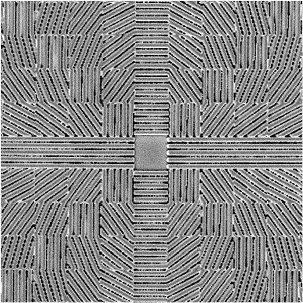
This scanning electron microscopy image shows microchannels fabricated by two-photon polymerization. Metin Sitti and co-workers used these microchannels to mold 3D liquid crystal network microstructures. These microchannels were designed using a “pixelated” technique with equally spaced, 10 μm × 10 μm pixels. This technique enables the fabrication of a variety of channel patterns, allowing more freedom when designing liquid crystal network microstructures.
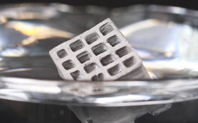
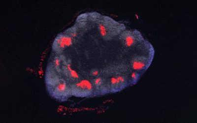
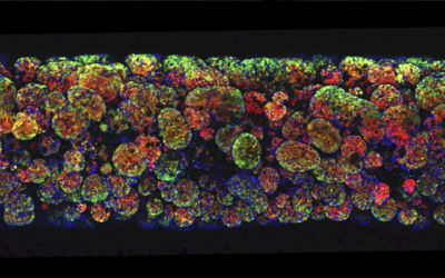
Kidney stem cells
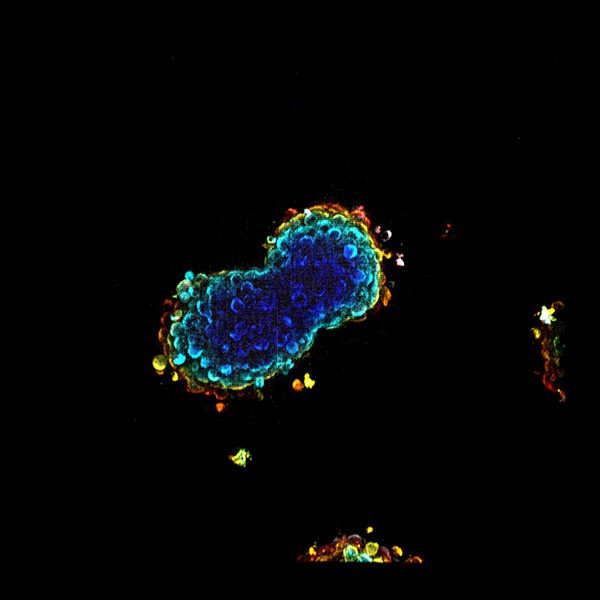
This spheroidally shaped cell culture of human embryonic kidney cells was templated by a photochemical technique. Vincent Studer and co-workers used photochemistry to controllably create hollow shapes within hydrogel structures. These hollows were then seeded with cells, which grew to fill the empty space. The spheroids were stained using a cell marker and the 3D imaging was accomplished with a bespoke digital micromirror-device-based confocal microscope.
Sneaky microrobots
Zwitterionic polymers allow researchers to fabricate drug-carrying microrobots that can operate covertly under the immune system’s radar. Salvador Borros, Metin Sitti, and their teams created these sneaky bots using fully zwitterionic photosensitive materials developed for two‐photon polymerization and 3D microprinting. The microbots have ample functionality, with tunable mechanical properties, anti‐biofouling and non‐immunogenic properties, functionalization for magnetic actuation, encapsulation of biomolecules, and surface functionalization for drug delivery.