")
In a captivating display of the seamless integration of art and science, these groundbreaking experimental images showcase the striking beauty hidden within the realm of scientific exploration. These images serve as a testament to the intricate relationship between artistic expression and scientific discovery.
From vibrant microscopic landscapes to mesmerizing patterns formed by intricate molecular structures, these visuals not only captivate the eye but also provide a glimpse into the fascinating world of scientific research. Each image tells a unique story, unveiling the wonders that lie beneath the surface of our understanding.
Scientists and researchers are increasingly recognizing the power of art to communicate complex scientific concepts and ignite curiosity in audiences. By carefully crafting these mesmerizing visuals, they are able to convey the profound beauty and significance of their discoveries, transcending disciplinary boundaries.
Through the lens of art, we gain a renewed appreciation for the remarkable achievements in scientific exploration. These captivating images allow us to delve deeper into the wonders of the natural world, reminding us that the pursuit of knowledge can be as visually breathtaking as it is intellectually enriching.
Feature image credit: Virgilio Mattoli, et al., published in Advanced Functional Materials.
The ultimate protein delivery vehicle
Coacervates — aqueous phases rich in macromolecules such as synthetic polymers, proteins, or nucleic acids — have revolutionized protein delivery, especially within the realm of tissue engineering and regenerative medicine. However, there’s been one obstacle standing in the way of its widespread use: a tendency to clump together.
In a study published in Macromolecular Bioscience, researchers led by Yadong Wang of Cornell University have developed a remarkable method to overcome this limitation. Through a cutting-edge process, phospholipids are assembled on the surface of the coacervate, resulting in the formation of LipCo (shown above), a discrete spherical structure, like a tiny fortress, with the coacervate residing inside and the phospholipid serving as its protective shield.
LipCo stands strong against the test of time, preserving the bioactivity of proteins and emerging as the ultimate protein delivery vehicle, armed with enhanced colloidal stability.
Unlocking the power of sound
Imagine having the ability to control sound waves with precision and finesse. Acoustic metasurfaces, an innovative technology, hold the key to this extraordinary capability.
Using an ultrathin metasurface approach, David Collins and his team at the University of Melbourne were able to create planarized micropillars arranged in a discrete phase array, forming a subwavelength metasurface. The best part? This metasurface can be effortlessly made through a single-step etching process, making it accessible and practical for a wide range of megahertz-scale applications.
In their study published in Advanced Materials, complex acoustic patterns come to life through the magic of acoustic holography. Harnessing the potential of this metasurface, the team demonstrated its ability to produce intricate and customized acoustic patterns, opening up a world of possibilities.
Nanobelt haystacks
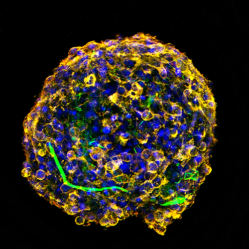
Overcoming the challenges of using neural stem cells to treat spinal cord injury has long been a quest in the field of regenerative medicine. In a groundbreaking study published in Advanced Functional Materials by Yuanhua Sang, Hengxing Zhou, Hong Liu, the team has devised a remarkable solution by engineering customized hybrid spheroids that possess a unique architecture, thanks to the incorporation of a specific nanobelt haystack framework.
This framework serves a dual purpose: providing structural support to alleviate hypoxia within the spheroid core and promoting the crucial biological process of neural differentiation. This approach not only offers a path for overcoming the limitations of stem cell therapy but also highlights the significant role of nanomaterials in facilitating cell-material interactions.
Shrinking the power of detection
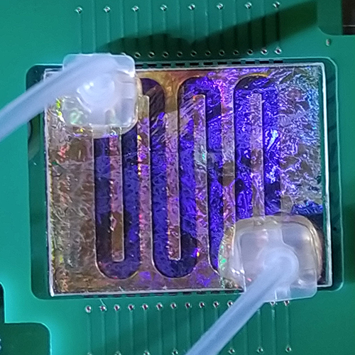
The era of miniaturized optical biosensors has arrived, heralding a new chapter in analysis and diagnostics! In a study published in Advanced Materials, scientists led by Margherita Bolognesi and Stefano Toffanin of the Institute of Nanostructured Materials (ISMN) – National Research Council (CNR) demonstrated a fully miniaturized optical biosensor prototype based on plasmonic detection, paving the way for fast and multichannel sensing of analytes with varying molecular weights.
The scientists demonstrated its capabilities in analyte-specific and rapid immunoassay-based detection, achieving results within a mere 15 minutes. With its ability to bring high-performance sensing into portable and real-world applications (such as testing the safety of milk), this technology has the potential to transform various fields, from food safety to medical diagnostics.
Rolled-up thin films
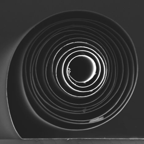
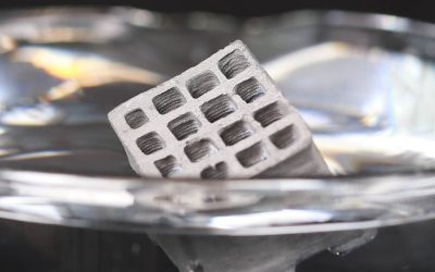
Imagine being able to transform a flat, thin film into a complex, 3D structure with just a single step. This technology, called rolling origami, has made it possible to create microstructures with remarkable precision and functionality. However, until now, constructing complex rolling structures in a high-yield and scalable manner has been quite challenging.
In a study published in Advanced Science, scientists led by Zaklina Burghard from the University of Stuttgart have devised a simple yet effective fabrication technique that uses external mechanical stress to roll micrometer-thick, flexible films into tubular or spiral shapes within seconds. The process is incredibly efficient and allows for precise control over the diameter of the rolled structures, the number of windings, and even the surface morphology at the nanostructural level.
This method can be applied to a wide range of materials with different functionalities. The resulting three-dimensional structures hold immense promise for various applications. For instance, they can be used as sensors to detect and measure physical or chemical changes in the environment.
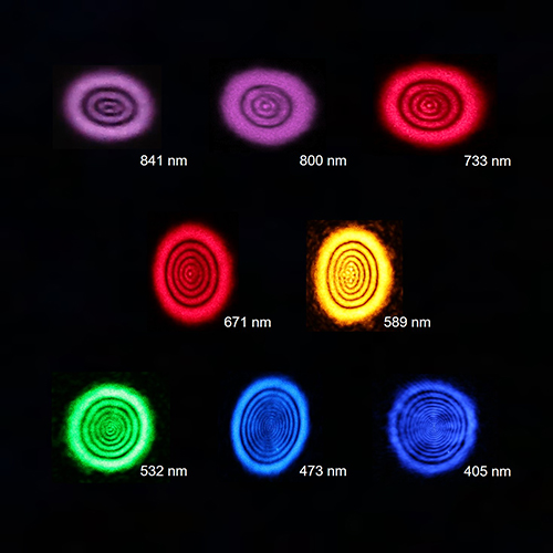
Elastic photonic shells
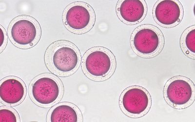
These are tiny particles called photonic microspheres that can be used to add color to inks and cosmetics. In their study published in Advanced Optical Materials, scientists led by Shin-Hyun Kim of KAIST created them with elastic photonic shells, lending stronger colors and greater flexibility.
They used a template that involved enclosing water droplets within oil droplets, which were then surrounded by another water layer. By using a certain type of resin and silica particles in the oil layer, the researchers were able to improve the color quality of the shells, and with a liquid core, they could change shape easily.
The color of the shells could be adjusted by changing the conditions they were exposed to. These elastic photonic shells offer exciting possibilities for enhancing the color and flexibility of inks and cosmetics.
Ships passing in the night
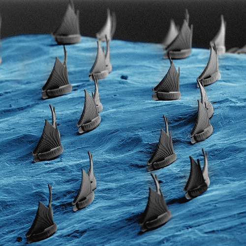
Fabricating micro- and nano-scale objects on complex surfaces is crucial for the advancement of cutting-edge technologies. This captivating image was produced by team led by Virgilio Mattoli at the Center for Materials Interfaces, Istituto Italiano di Tecnologia, Italy and published in Advanced Functional Materials.
Employing a two-photon polymerization printing technique, the team successfully crafted these awe-inspiring ships braving a stormy sea. Utilizing ultra-thin polymeric films as a supportive medium, the printed micro-structures are suspended and transferred onto the designated surface with precision. Once the process is complete, the nanofilm can be effortlessly removed, except for the area beneath the printed elements, where it acts as a thin adhesive layer measuring tens of nanometers.
The visionary creations brought to life by this team also include a captivating “micro diver” (see this article’s feature image) and intricately patterned geometric surfaces.
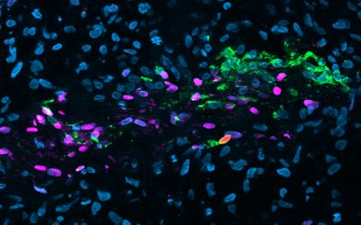
The role of astrocytes
Astrocytes play an important role in maintaining a healthy brain environment by regulating various factors like ions, water, and neurotransmitters. They rely on the dynamic behavior of the actin cytoskeleton, particularly near the cell boundary, to regulate homeostasis in response to specific environmental cues. The topography of the cell’s surroundings and interactions with neurons further influence the dynamics of the cytoskeleton.
This image taken from the study published in Advanced Biology by Valentina Benfenati of the National Research Council of Italy, Wolfgang Losert from the University of Maryland, and their teams showcases a STED microscopy image of the nanoscale actin structure within primary astrocytes, providing valuable insights into the active role of astrocytes in the functioning of neural networks.
Responsive fabrics
Smart textiles that can automatically respond to environmental changes are highly sought after, and liquid crystal elastomers (LCEs) have shown promise as base materials.
In a study published in Advanced Materials, researchers led by Jaana Vapaavuori of Aalto University and Eugene M. Terentjev of Cambridge University have successfully crafted active fabrics by incorporating LCE yarn into woven structures using a standard loom. They tested two types of LCE yarns, one soft and one stiff, and explored various weaving patterns, identifying two standout performers: the twill pattern with stiffer LCE yarn, which exhibited the highest blocking force, and the weft rib pattern, which showed over 10% reversible actuation strain during repeated heating cycles.
The active fabrics created using LCE yarns demonstrated reversible three-dimensional shape changes by utilizing circular weaving patterns that resulted in cone shapes when heated. This seamless integration of active LCE yarns with existing passive yarns opens up new possibilities for developing stimuli-responsive textiles that can actuate and respond to different stimuli.
A walking ferroelectric liquid droplet
Researchers have discovered a fascinating phenomenon involving the motion of ferroelectric liquid droplets on a ferroelectric lithium niobate substrate. In their study published in Advanced Materials, a team led by Tommaso Giovanni Bellini of the University of Milano and Liana Lucchetti of the Università Politecnica delle Marche found that by illuminating the substrate with a moderate-intensity light beam positioned a few droplet diameters away, they were able to control the movement of the droplets.
Remarkably, the researchers found that they could manipulate the droplets over long distances by moving the light beam. This behavior is attributed to the interaction between the polarization of the ferroelectric droplets and the polarization induced in the irradiated region of the lithium niobate substrate.
These findings shed light on the complex interplay between polarization and light in ferroelectric liquid crystals, opening up new possibilities for the development of advanced light-driven actuators and other functional devices.
Quantum materials
Manipulating quantum materials through laser-induced electron coherence has attracted significant interest. A recent study published in Advanced Materials by Jimin Zhao fro the Chinese Academy of Sciences and collaborators has demonstrated that this phenomenon in 2D materials can lead to a unique nonlinear optical response called spatial self-phase modulation (SSPM), enabling the development of a novel all-optical switching method. However, most investigations have focused on electron coherence, neglecting the exploration of laser-induced hole coherence.
In this study, researchers have observed the optical Kerr effect, a nonlinear phenomenon, in flakes of a 3D material called Weyl semimetal TaAs. These observations provide valuable insights into the behavior of quantum materials under laser-induced coherence and offer potential avenues for developing new applications in nonlinear optics and quantum information processing.
The study highlights the importance of considering both electron and hole coherence in the exploration and manipulation of quantum materials.
Networks of light-sensitive nanowires
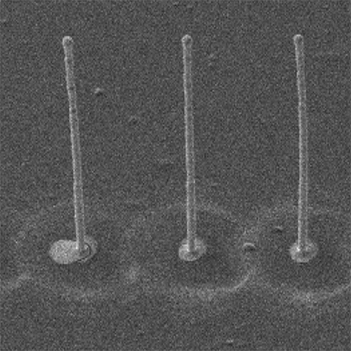
When it comes to creating intricate, detailed features on a tiny scale, additive manufacturing (or 3D printing) often falls short. In this study by Ravinder Dahiya from Northeastern University and team, the scientists have found a way to overcome this limitation by optimizing a special printing technique called drop-on-demand (DoD) electrohydrodynamic (EHD)-based jet printing.
They used this method to create tiny pillars made of gold (Au), measuring less than the width of a human hair. These micropillars were arranged in arrays, forming electrode structures. But the innovation doesn’t stop there. By combining AM with a process called hydrothermal growth, the researchers achieved something truly remarkable. They were able to grow zinc oxide (ZnO) nanowires directly on the printed gold micropillars, without needing any additional seed material. This hybrid approach resulted in the formation of intricate networks of light-sensitive nanowires.