“Art and science have their meeting point in method” – Edward Bulwer-Lytton. This series of photographs published recently in our journals features incredible images which caught the eye of our editors.
Little big hearts
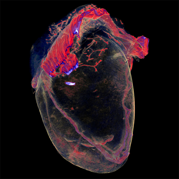
This micro-CT image of a mouse heart forms the basis of the research by Pierre Weiss at the University of Nantes and his team, wherein in situ formation of injected hydrogels are used for tissue engineering applications. The gels show good biocompatibility and degradation in vivo with tunable control over the hydrogel’s mechanical properties. This stunning image deserved the front cover of issue 19 of Advanced Healthcare Materials.
Stunning nano-flowers
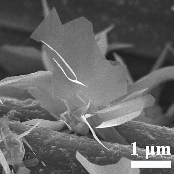
This image published in Advanced Materials Interfaces by a collaborative group of researchers demonstrates the controlled growth of rhenium disulfide (ReS2) nanoflowers using ambient pressure chemical vapor deposition. ReS2 shows great promise as an electrocatalyst for the evolution of hydrogen, and methodologies that can fine-tune it’s surface have the potential to propel its catalytic capabilities.
Purple haze
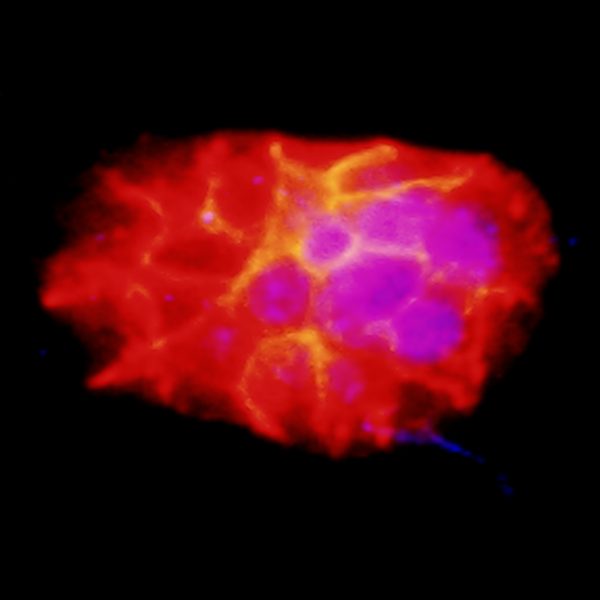
In work published in Advanced Functional Materials, keratin cells are treated with “carbon dots”. The blue stain shows the cell nuclei and the red shows cell markers presented on the cell membrane. The carbon dots stimulate a signalling pathway that leads to more cell movement and faster healing of wounds in mice.
Lotus leaf mimic
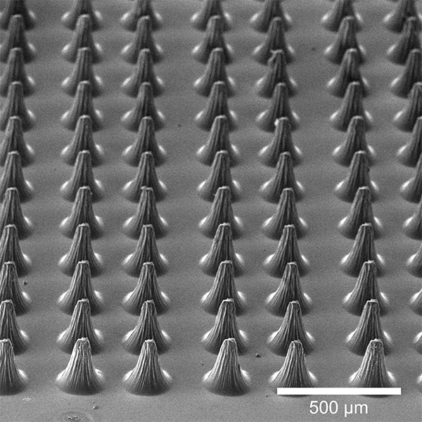
The micropapilla on the surface of lotus leaves help keep them clean and contribute to structural integrity. In Advanced Materials Interfaces, Lei Wang and team show that synthetically manufactured micropapilla can prevent material rupture. These synthetic micropapilla are made from a 1mm thick sheet of silicone and the spacing between the pillars can be optimized to give improved material durability.
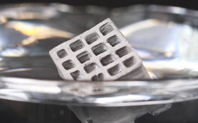
A transformable tube array
Never mind 3D printing, in Advanced Materials Lee and Lee report a 4D printed transformable tube array that can directly transfer a large number of 3D culture models from a microwell plate to a much smaller histology cassette. While seemingly innocuous, this new device is actually revolutionary for high throughput 3D culture analysis as it allows researchers to quickly assess the internal microstructure of 3D cell cultures without the need for manual, laborious, and time‐consuming histological procedures.
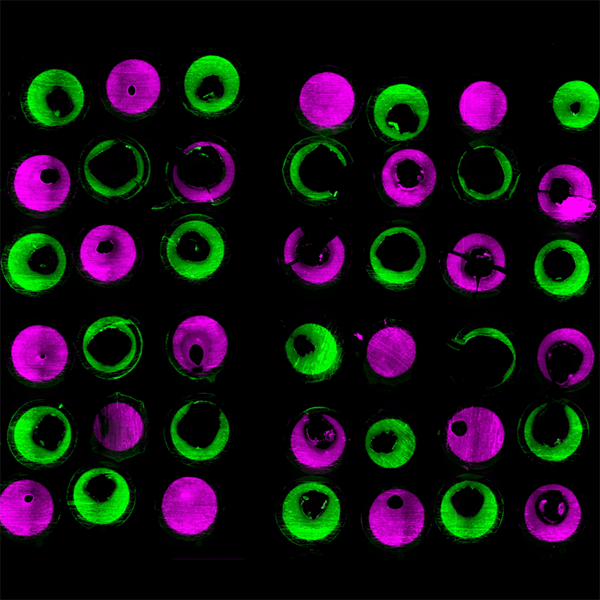
Galactic clouds or organ model?
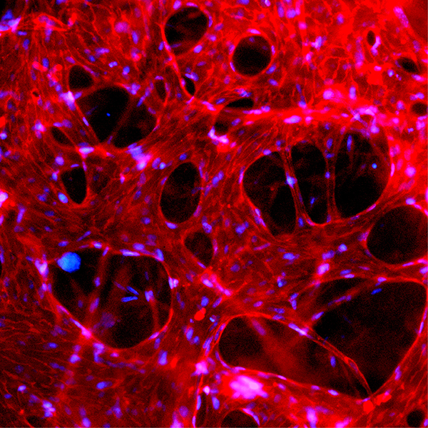
3D organoids are miniaturized models of organs, but their lack of vascularization — or ability to develop blood vessels — limits their ability to effectively mimic physiology. In Advanced Materials, Boyang Zhang and co-workers develop an innovative microfluidic platform called IFlowPlate, which can be used to culture up to 128 independently perfused and vascularized colon organoids, unlocking new possibilities for screening therapeutics or modeling relevant diseases.
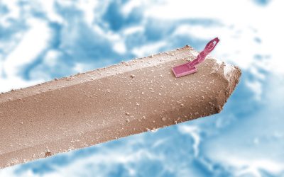
Are you sure these are your measurements?

Well, ladybirds get married too right? This ring has a diameter of around 15 mm. Published in Advanced Materials, it was made as part of research looking into a special 3D printing technique. In this protocol, a projection of the 3D image is cast in UV light on a bulk liquid resin. The sections of resin that are sufficiently exposed to this light cure into a solid while the rest of the bulk stays in the liquid phase. This allows the ready preparation of 3D objects that are free from layering defects.
Patterned rose petals
This mesmerizing image actually depicts patterns of particles sintered onto a delicate rose petal. Given that metals and organic materials have different surface energies, bonding them together has posed quite a challenge, which is especially problematic when it comes to developing wearable devices. Through a new universal metal printing method reported in Advanced Materials Interfaces, Martin Thuo and coworkers demonstrate the ability to print metal designs onto biological tissues (like the brain!) and organic substrates.
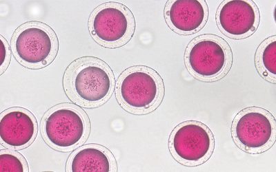
Fluorescent microgels
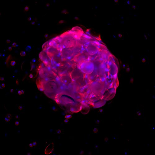
While this looks like a shot out of Stranger Things, this image by Careia, Mano, and co-workers captures a fluorescent microgel that encapsulates living cells for tissue reconstruction. Their study published in Advanced Biosystems describes the high throughput fabrication of liquefied capsules, built using microarrays to produce the initial structures for tissue engineering.
See more in this series “Science in pictures“