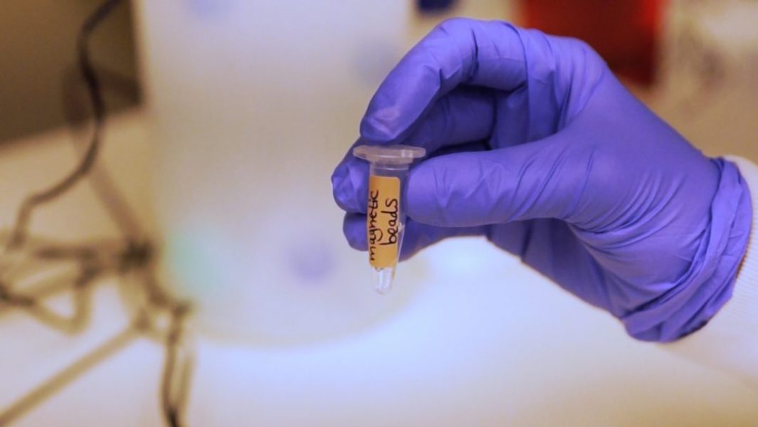
Clinical research and diagnostics frequently make use of fluorescence‐based immunoassays due to their high sensitivity and ease of operation. However, the autofluorescence of capture surfaces, such as magnetic beads, produce strong background noise.
In Small, Dr. Amos Danielli from Bar‐Ilan University, Israel and co-workers devise a novel photobleaching method to reduce the autofluorescence of magnetic beads, improving the analytical performance of fluorescence‐based immunoassays.
Dr. Amos Danielli: The system we use to photobleach the beads includes a laser that is reshaped by two planar convex lenses, is reflected by a dichroic beamsplitter, and focused by an objective lens onto the sample of magnetic beads. Two electomagnets aggregate the magnetic beads to one side of the cuvette, and the laser excites the magnetic beads, and the emitted autofluorescence is detected by a photomultiplier tube and displayed on the oscilloscope.
Shira Roth: The autofluorescence properties and the stability of the photobleaching were analyzed over a period of two months, indicating that the photobleached beads were stable over time and their surface functionality was retained.
A commercially available immunoassay, Human Interleukin-8, was detected with a three-fold improvement in the detection limit and signal-to-noise ratio of immunoassays compared to non-bleached beads, and autofluorescence was reduced to 1% of the initial value.
To find out more about this photobleaching method to reduce the autofluorescence of magnetic beads, please visit the Small homepage.

