The interaction of biological fluids with nanoparticles (NPs) upon in vivo injection determines the NPs bio-identity due to the formation of a coating on their surface. This coating known as a biomolecular or protein corona is made up of many different components, and can negatively affect the therapeutic competency of the NPs. Serum proteins, lipids, and small metabolites adsorb to the NPs surface affecting their stability, targeting ability and immunogenicity.
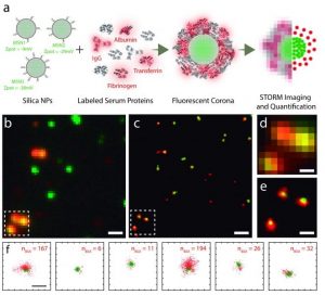
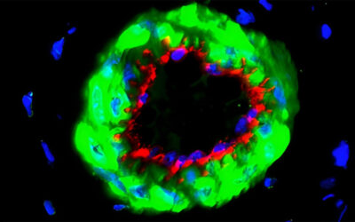
STORM imaging of protein corona formation in mesoporous silica nanoparticles. Nanoparticles are shown in green and the labeled protein in red. More information here.
An understanding of the formation and dynamics of this coating is needed to enable the effective design of nanostructures for use in biomedical applications. Some studies have focused on ex situ and in situ research of the corona; however, the study of NP-protein interaction is challenging due to the complex nature of the serum and the small size of the NPs. Therefore, advanced techniques are required to study the corona composition for biomedical nanomaterials.
In recently published work, Lorenzo Albertazzi and co-workers, look to provide a greater understanding of the corona formation on NPs with the use of stochastic optical reconstruction microscopy (STORM) as a single molecule localization microscopy technique. STORM provided excellent spatial resolution, molecular specificity, and single molecule sensitivity. This super-resolution optical microscopy technique enabled the imaging of the heterogeneity and the dynamics of the NP protein corona.
While STORM has previously been used in cell biology, only recently has it be employed in the field of nanotechnology. Direct STORM (dSTORM) was employed in this recent work to investigate the interaction between silica NPs and serum proteins, and provided nanometric accuracy at the single particle level.
The relevance of the corona formation in NP cancer cell targeting is shown, instilling the importance of understanding such phenomena when designing NPs for targeting applications. Super-resolution microscopy has broad and latent application for various nanomaterials, representing a tool for the study of protein corona formation, even in physiological conditions under flow, and in human patients’ plasma.
For more information about this technique and the recent work from Lorenzo Albertazzi and co-workers, please refer to original manuscript in Small.