
A flexible wide-field endoscope utilising blue excitation light provides label-free contrast of tissue based on fluorescence lifetime imaging of tissue autofluorescence.
 Fluorescence lifetime imaging (FLIM) of tissue autofluorescence has been shown to provide labelfree contrast between different types and states of tissue. Clinical FLIM can combine lifetime contrast with morphological information to provide a direct comparison between different spatial regions – making it easier to spot differences with respect to “normal” tissue, e.g. for diagnostic screening and potentially enabling margins of diseased tissue to be identified. To date, however, there have been relatively few clinical FLIM studies, partly due to a lack of suitable instrumentation.
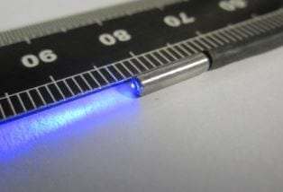
With the aim of applying FLIM to detect and monitor disease in internal organs such as the colon, a British team led by Hugh Sparks (Imperial College London) has now developed a flexible wide-field FLIM endoscope based on coherent optical fibre bundles. The device utilizes low average power blue picosecond laser diode excitation sources to induce tissue autofluorescence. Its multimode optical fibre efficiently delivers the excitation radiation to illuminate a 3 mm field of view.
Unfortunately, the multimodal optical fibre propagation necessary to achieve this broad illumination can lead to errors in the lifetime determination due to temporal broadening of the excitation pulses. To address this issue, the researchers characterized the consequent degradation of the spatio-temporal instrument response function (IRF) and incorporated a spatially varying temporal instrument response function in the FLIM analysis.
As prove of principle, the scientists applied their new FLIM endoscope to ex vivo tissue autofluorescence from diseased human larynx biopsies. Using a gain-switched picosecond diode laser operating at 445 nm as the excitation source and an average excitation power of circa 0.5 mW, mm-scale spatial maps of autofluorescence lifetimes could be acquired in about 8 seconds.
To illustrate the instrument’s potential for FLIM at higher acquisition rates, a higher power mode-locked frequency doubled Ti:Sapphire laser was used to carry out FLIM of ex vivo mouse bowel at up to 2.5 Hz using 10 mW of average excitation power at the specimen.
The results demonstrate the potential of the technique to screen for neoplasia. Based on the flexible wide-field FLIM endoscope, new clinical instrumentation may be developed to aid diagnosis and monitoring of therapeutic interventions.


