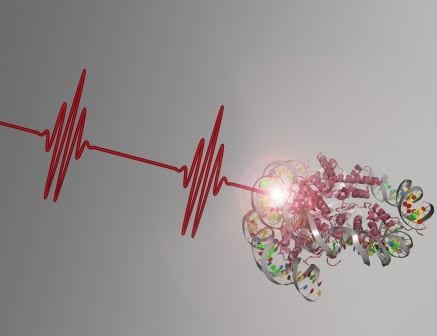
Nonlinear photoperturbation of chromatin with femtosecond laser pulses.
A new straightforward method enables monitoring the response of nuclear proteins to DNA damage in time and space. The approach is based on nonlinear photoperturbation.
Understanding the cellular response to DNA strand breaks is crucial to decipher the mechanisms maintaining the integrity of our genome. In eukaryotic cells, the molecular events triggered by DNA damage are strongly influenced by the local chromatin environment surrounding the lesion. The ensuing DNA repair process, in turn, impacts on chromatin structure. In order to understand how these two fundamental processes are mutually connected, both chromatin rearrangements induced by DNA damage as well as DNA repair activity have to be visualized in living cells.
Elisa Ferrando-May and a team from University of Konstanz now present a novel method to visualize how the mobility of nuclear proteins changes in response to localized DNA damage. Their new approach enables to inflict DNA damage without interfering with a subsequent mobility measurement by fluorescence photoactivation. It is based on nonlinear photoperturbation using infrared femtosecond (fs) laser pulses. The assay detects how the dynamics of nuclear proteins is affected by localized DNA strand breaks, irrespective of their recruitment behavior.
The scientists induce DNA strand breaks via nonlinear excitation with fs laser pulses at 1050 nm in a 3D-confined subnuclear volume. After a time delay of choice, they analyze protein mobility within this volume by two-photon photoactivation of PA-GFP fusion proteins at 775 nm. The time delay can be chosen freely permitting to probe protein mobilities at different subsequent stages of the DNA damage response. By changing the position of the photoactivation spot with respect to the zone of lesion the influence of chromatin structure and of the distance from damage can be investigated. Due to the local confinement of multiphoton absorption it is possible to examine spatially distinct subnuclear areas, such as eu- and heterochromatin.
As first applications, the scientists demonstrated a locally confined, time-dependent mobility increase of histone H1.2, and a progressive retardation of the DNA repair factor XRCC1 at damaged sites.


