From molecular structure to tissue architecture: second-harmonic generation (SHG) microscopy is an ideal tool for probing collagen organization. For example, it can be used to study skin tumors and skin ageing.
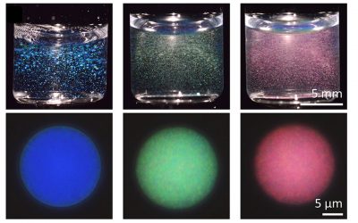
Second-harmonic generation (SHG) is a nonlinear second order optical process occurring in noncentrosymmetric systems with a large hyperpolarizability. This condition is satisfied at the molecular level by the presence of electron-donor and electronacceptor moieties connected by a π-conjugated system and by a structural organization of the molecular emitters at the focal volume scale. Both conditions are satisfied in anisotropic biological molecules such as collagen, making SHG microscopy an ideal tool for imaging collagen and probing its hierarchical organization from molecular scale up to tissue architectural level. SHG combines the advantages of a non-linear microscopy approach with a coherent modality able to probe molecular organization.
In a review article, Riccardo Cicchi from European Laboratory for Non-linear Spectroscopy (LENS), in Sesto Fiorentino, Italy, and his co-authors from Italy and Jena, Germany, sum up the physical concepts of SHG from collagen. They explain how this optical process allows probing structures ranging from molecular sizes to tissue architecture, through image pattern analysis and scoring methods. Starting from the description of the most relevant approaches employing SHG polarization anisotropy and forward – backward SHG detection, the scientists then focus on the most relevant methods for imaging and characterizing collagen organization in tissues through image pattern analysis methods. They outline the advantages and limitations of the methods applied to tissue imaging as well as potential clinical applications.
SHG has already been used for imaging collagen rich tissues such as cornea, tendon, and arteries. In particular, SHG microscopy has been mainly used for selectively investigating collagen fibres orientation and their structural changes in human dermis, keloid, fibrosis, thermally-treated samples, and also in tumor microenvironments.
The Second-harmonic to Autofluorescence Ageing Index of Dermis (SAAID) is a simple scoring method to be used when photon excited fluorescence microscopy (TPEF) and SHG microscopy are used simultaneously for imaging connective tissue. Since collagen and elastic fibres can be respectively imaged by SHG and TPEF microscopy, this method can be used not only to determine ageing but also other altered physiological conditions of skin dermis and, more in general, of connective tissue. It can be used to study skin tumors.