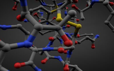
South Korean researchers have developed an improved technique for high-resolution fluorescence imaging of biomolecular events taking place in cells on surfaces. They used it to observe an adenovirus being taken up by cancer cells, which cannot be watched easily using conventional methods.
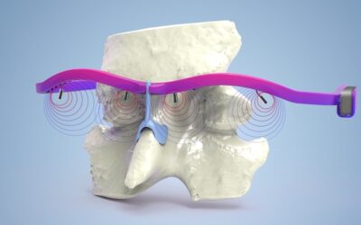
To study the processes and movements of cells, it is important to get clearer and more detailed pictures of them. Many methods are currently used to try to achieve this, but they often do not have good resolution in both the horizontal and vertical directions. Some methods require the production and presence of specific nanostructures on a surface which enhance resolution near to the nanostructure only; these specific nanostructures are not straightforward to produce reliably on a large scale, which would be necessary for everyday use in a laboratory. One technique, total internal reflection spectroscopy (TIRF) has previously been combined with other techniques to give imaging higher resolution. TIRF works by measuring generated electromagnetic waves that disappear exponentially, producing fluorescence only on the surface of an object. This effect is called evanescence.
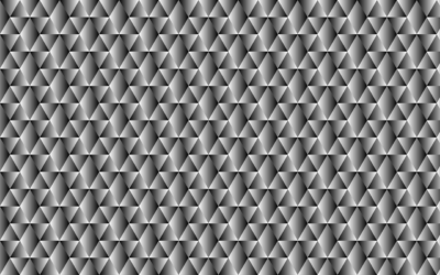
In this study, Chae-Ok Yun, Donghyun Kim and co-workers at Yonsei University, South Korea, created tiny gold or silver nanoislands on a surface and employed these to generate very small hotspots of evanescence on the surface, which were used to create an image by TIRF. The nanoislands are easy to make in a controllable way, and the geometry, size, and distribution of the nanoislands determine the resolution of the resulting images.
The researchers tested their improved technique, christened SUPRA-TIRF for surface-plasmon-enhanced randomly activated TIRF, on two types of cancer cells that were taking up fluorescent beads and adenoviruses by endocytosis. They were able to watch the cells swelling after injection of the virus. This new technique allowed the researchers to visualise a single adenovirus particle, which is too detailed for conventional TIRF to see. The measured fluorescence intensity of the beads was eight times higher for SUPRA-TIRF than when using regular TIRF, which makes the new system much more sensitive.
The technique could in the future be an invaluable tool for visualisation of other cells and cellular events, thereby aiding in biomedical research of many different sorts. These studies of cancer cells are just the beginning.