
Infamously delicate work, the surgical removal of tumours from the brain is complicated by the interpenetrating nature of the tumour tissue with the healthy surrounding tissue.
Research from Fudan University, China, could now allow brain surgeons to identify malignant tumour tissue more easily, with the help of an old favourite: gold.
They have developed two sets of probes made of biocompatible gold nanospheres functionalised with either alkynes or azides, and protected by a shielding layer.
When injected intravenously, the probes are able to cross the blood–brain barrier. Encountering normal, healthy brain tissue, they remain intact and make their way back into the bloodstream, from which they can be safely and rapidly excreted.
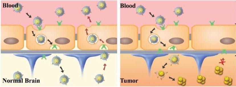
When the probes find themselves in the harsher environment of a tumour, however, the increased acidity removes the shielding layer and allows the azide and alkyne functionalities to react, forming bulky aggregates through click cycloadditions.
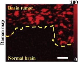
These aggregates stick in the tumour tissue and can be detected due to their activated signals either via pre-operative magnetic resonance imaging or surface-enhanced resonance Raman scattering (SERRS). The particular advantage of the SERRS technique is the availability of hand-held Raman scanners, which can be used in the operating room, effectively with the scalpel in the other hand.
These pH responsive nanoprobes are expected to greatly improve brain tumour surgery outcomes by allowing the surgeons to clearly define the boundaries of tumour tissues.
See further details in Advanced Materials.


