
Lymphatic malformations (LM) are complex congenital vascular lesions composed of dilated, abnormal lymphatic channels of varying size that can result in significant esthetic and physical impairment due to relentless growth. Lymphatic malformations comprised of micro‐lymphatic channels (microcystic) integrate and infiltrate normal soft tissue, leading to a locally invasive mass. Around 1-in-2000 to 4000 babies are born with lymphatic malformations and the disease management is a challenge due to several factors, one being diagnosis.
Clinical history, physical exam, and radiographic imaging are used in the diagnosis of LM. Imaging techniques like ultrasonography and magnetic resonance imaging assist in the diagnosis but are unable to detect microvasculature present in microcystic lymphatic malformations. For example, ultrasonography has poor spatial resolution and is not effective for detailed microvascular imaging. But better diagnose alternatives are in the making.
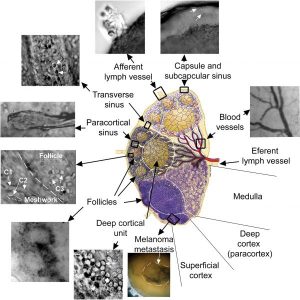
Optical imaging of microanatomy of the fresh mouse lymph node ex vivo obtained with TOC using 80% glycerol water solution
A group of Russian and American researchers examined existing tools and elaborated on alternative diagnostic methods in assessing lymphatic malformations in a new review article including photoacoustics.
Photoacoustic technology is one of the fastest growing research areas in biomedicine. PA‐based methods, which are based on the nonradiative relaxation of absorbed energy (e.g., a photon is not emitted) into heat and then into sound, offer an advanced combination of parameters beyond the capability of many other optical methods including (1) insensitivity to light scattering and autofluorescence, (2) the ability to assess deep (up to 3‐5 cm) tissue structures, and (3) clinical applicability with safe laser energy exposure levels. In preclinical (in vivo animal models) and clinical studies, PA methods have already demonstrated great promise for advanced diagnosis of different diseases.
In particular, photoacoustics, low‐toxicity nanoparticles and optical clearing can overcome existing challenges in the examination of lymphatic channels in vivo. In combination with photothermal scanning and flow cytometry, photoacoustic techniques may provide a versatile tool for lymphatic‐related clinical applications, potentially leading to a single diagnostic and therapeutic platform to overcome limitations in current imaging techniques and permit targeted theranostics of microcystic lymphatic malformations.
The advantage of the described technology is that it involves safe laser parameters and low‐toxicity molecular nanoprobes, has high potential for use with human subjects to overcome the limitations of current imaging techniques, and permits targeted theranostics of microcystic LM. Additional diagnostic improvement can be achieved through integration of established and novel methods such as different combinations of US, MRI, X‐ray, and PA technology.


