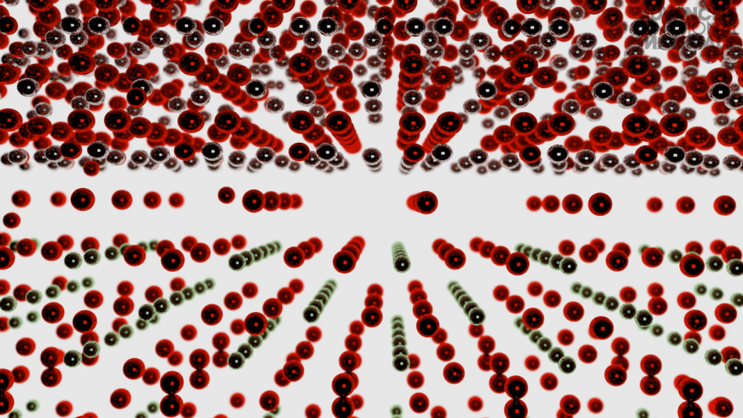
Oxide electronics, photovoltaics, and catalysis are just some of the practical applications of functional oxides. Understanding and predicting the defect structure and formation in complex oxides allows us to precisely design oxide-based nanodevices for unique functional applications.
In their paper in Advanced Functional Materials, Felip Sandiumange and colleagues from the Institut de Ciència de Materials de Barcelona (ICMAB-CSIC), and co-workers from the Catalan Institute of Nanoscience and Nanotechnology (ICN2), Ohio State University, and the University of Belgrade, combine atomic resolution imaging and spectroscopic techniques to determine the complex structure of misfit dislocations in a perovskite-type heteroepitaxial system (La0.67Sr0.33MnO3/LaAlO3).
Dr. Felip Sandiumenge: “The dislocation strain field in oxides may couple strongly with the defect chemistry of the host crystal, and, as a consequence, the physical properties of dislocations in oxides may differ substantially from those of the material which contains them. This behavior, in fact, offers interesting opportunities because it allows to envisage nanodevices inspired on self-organization of naturally occurring defects in crystals.”
Dr. Núria Bagués: “In this work, we combine high-resolution transmission electron microscopy (HRTEM) and high-angle angular annular dark field (HAADF) to determine the structure of the dislocation core.”
HAADF images, sensitive to the atomic number, reveal the atomic structure of the dislocation core. Complementary HRTEM images, on the other hand, are used to map the dislocation strain field.
Dr. Núria Bagués: “In addition, we use energy dispersive X-ray (EDX) spectroscopy to map the cation distribution and the oxygen vacancies over the strain field, and electron energy loss spectroscopy (EELS) is used to determine the oxidation state of the manganese cations.”
EDX mapping and EEL spectra across the dislocation core show depletion of oxygen atoms and reduction of Mn cations to Mn3+ around the tensile region of the dislocation strain field. This results in electrostatic interactions that control the cationic distribution around the dislocation core.
To find out more about misfit dislocations in this perovskite-type heteroepitaxial system, please visit the Advanced Functional Materials homepage.


