Quantum cascade lasers enable the quick and efficient real-time mid-infrared microspectroscopy of living microorganisms. A slowly moving amoeba and a quick moving nematode were imaged to demonstrate the performance of the method.
 The combination of microscopy with spectroscopy is frequently referred to as ‘hyperspectral imaging’ – also called ‘chemical imaging’ if molecular vibrations by means of mid-infrared (MIR) or Raman spectroscopy are considered. This non-destructive, reagent- free method has become an important tool for the structural and spectral analysis of biological samples in vitro.
Initially, MIR microspectroscopy was based on Fourier-transform infrared mapping. Despite its successes, this method has some major drawbacks with respect to clinical applicability: long acquisition times, a spectral power density of the radiation source being too low to enable investigation of aqueous systems within a reasonable time, and the necessity of liquid nitrogen cooling of the detector. The invention of quantum cascade lasers (QCLs) opened up space for new opportunities in biomedical vibrational spectroscopy: The investigation of aqueous samples thicker than a few micrometers and of fast changing systems, such as living organisms.
In a new study, researchers from Heidelberg University (Germany) now demonstrate the great potential of QCL-based microspectroscopy to reach these goals. As model organisms, they used a slowly and a fast moving species: Amoeba proteus and Caenorhabditis elegans. The scientists were able to achieve acquisition frame rates high enough to monitor the living organisms while they move.
Key to success was the unique combination of a tunable QCL with a pixel-rich microbolometer array detector. The researchers reduced the frequency bandwidth to a desired narrow spectral region. By doing so, they could achieve frame rates of up to 50 Hz.
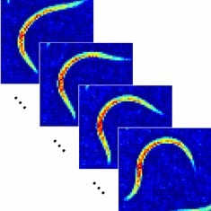
For the slowly moving amoeba, the laser was tuned over the wavelength range of 7.6 μm to 8.6 μm (wavenumbers 1320 cm–1 and 1160 cm–1, respectively). The recording of a hyperspectral image took 11.3 s whereby an average signal-to-noise ratio of 29 was achieved. The limits of time resolution were tested by imaging the fast moving nematode. The researchers chose a discrete wavenumber of 1265 cm–1 and performed mid-infrared imaging with the 640 × 480 pixel video graphics array (VGA) standard and at a full-frame time resolution of 0.02 s. This is well above the most common frame rate standards and represents, according to the authors, the first mid-infrared imaging of living organisms at VGA standard and video frame rate. An average signal-to-noise ratio of 16 was obtained in these experiments. (K. Maedefessel-Herrmann)
See the original publication: Katharina Haase, Niels Kröger-Lui, Annemarie Pucci, Arthur Schönhals and Wolfgang Petrich, Real-time mid-infrared imaging of living microorganisms, J. Biophotonics 9:1-2, 61-66 (2016)