The introduction of national screening programs to detect (pre-)cancerous processes, such as the Papanicoloau (‘Pap’) test and mammogram, have achieved considerable success and are estimated to prevent, for example, 4,500 and 1,300 deaths in the UK every year from cervical and breast cancer, respectively.
However, these programs require large, sustained financial input and human resources, and are not completely accurate. The ability to provide quantitative, objective and automated pathological analysis would provide enormous benefits for national cancer screening programs, in terms of both resource reduction and improved patient well being.
One technique with the potential to deliver a broader scope and automated classification is Raman spectroscopy. Raman spectroscopy is a label-free technique based on the inelastic scattering of light.
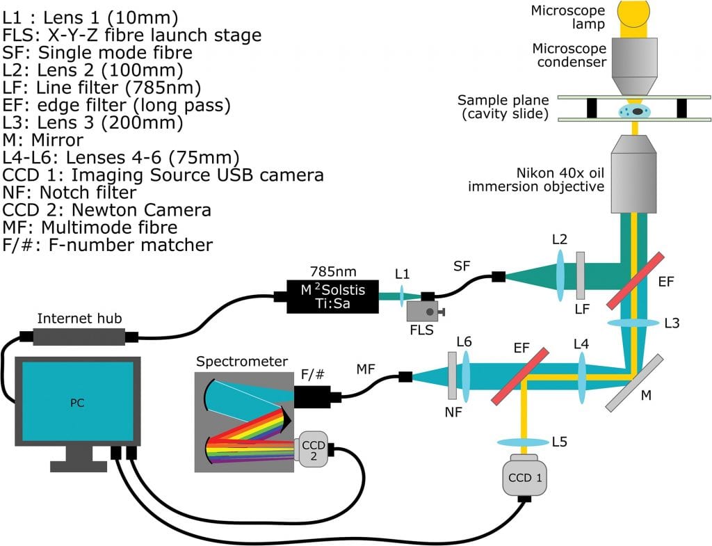
The optical diagram of the spectrometer designed for WMRS and standard Raman spectral acquisition. Brightfield microscope images are acquired separately on CCD1 and blocked from the spectrometer by secondary EF.
One of the major technical barriers to the introduction of Raman spectroscopy as a clinical tool is the long acquisition time associated with the rare event of Raman scattering of photons. The acquisition of high-quality spectra is further compounded by the presence of autofluorescence in samples, which is a particular problem for biological interrogation and can often lead to the loss of smaller Raman peaks obscured by a fluorescent background.
A team of researchers from the Edinburgh Cancer Research Centre and the University of St. Andrews in the UK tackled the problem. In their latest research they demonstrate the use of wavelength modulated Raman spectroscopy (WMRS) as a tool for the automated classification of unlabeled fixed cells in suspension, reflecting its potential utility in diagnosing clinical liquid based cytology samples.
WMRS is a wavelength-domain approach. During measurement, the laser wavelength is modulated throughout the acquisition process using a tunable laser.
“It is entirely feasible that the technique could be combined with other imaging methods such as Optical Coherence Tomography or Digital Holographic Microscopy in order to provide a cell topography map for automated point sampling”, according to team member C. Herrington.
It is hoped that such a system could be introduced to clinical settings in order to reduce the burden on manual cytological diagnosis, as well as increasing the likelihood of a correct outcome for patients.