In vitro drug sensitivity tests are usually performed in a monolayer, i.e. a two-dimensional (2D), culture system, which presents a largely artificial cellular environment with little relevance to in vivo three-dimensional (3D) conditions. These 2D systems have only limited value in predicting clinical efficacy of chemotherapeutic drugs. Conversely, 3D cell culture systems have great potential for cell therapy, tissue engineering and drug sensitivity testing because cells cultured as spheroids are more reflective of the in vivo microenvironment.
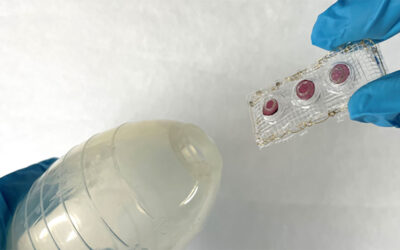
A recent study in Advanced Healthcare Materials reports the use of novel liquid marbles with high transparency and gas permeability for 3D cell culture and drug sensitivity tests of tumor spheroids. These liquid marbles were formed by gently rolling water droplets or a cell suspension on a bed of fume silica nanoparticles. These hydrophobic nanoparticles then automatically assemble on the air-liquid interface and help the droplets to form a spherical marble. With the force of gravity and internal flow the suspending cells in the liquid marbles aggregate at the bottom and grow into spheroids.
For the first time, the process of spheroid formation from single cells or a number of cells in liquid marbles can be recorded due to the high transparency of the material. In a subsequent drug sensitivity test the 3D spheroids show enhanced drug resistance compared to cell monolayers. The liquid marble technique may be further applied to create transparent spheroids from primary tumor cells isolated from clinical samples in order to select the most appropriate drugs for precision cancer therapy.


