The ability to measure blood flow in a noninvasive manner with high sensitivity and accuracy is of fundamental importance for studying a wide range of biological processes including local metabolism, neurovascular communication, pharmacodynamics, and stroke recovery.
Commonly used imaging modalities, such as magnetic resonance imaging (MRI), computed tomography (CT), and ultrasound (US) enable imaging of blood flow, but each of these methods has its downsides. Optical methods, on the other hand, can potentially be inexpensive, safe, while providing high throughput and ultimate sensitivity since a displacement in the order of the wavelength of the light can potentially be detected.
Among these techniques, photoacoustics (PA) is being intensely investigated for blood flow measurements due to its advantageous contrast mechanism based on absorption, and because of the relatively long wavelength of the ultrasound the technique is insensitive to small displacements. Ultrasound modulated optical tomography (UOT) was originally developed to image absorbing objects inside turbid media, and it has recently found exciting new applications such as focusing light into turbid media, measuring optical fluence variations, and quantification of scattering inhomogeneities. These applications of UOT are mainly motivated by the invention of a heterodyne parallel speckle detection (HPSD) method using a CCD camera.
A team of scientists from the MIRA Institute for Biomedical Technology and Technical Medicine in the Netherlands recently conducted a study where they used UOT with HPSD for imaging depth-resolved blood flow inside highly scattering media. They experimentally shown the dependence of the HPSD signal on the speed of blood flowing through a tube embedded in a scattering medium. The team also created an analytical model that relates the HPSD signal to the optical properties of moving particles and the background medium.
Their results demonstrate depth resolved imaging of blood in highly scattering media based on UOT using a HPSD method. “We demonstrated through a simple experiment that the ratio of the two UOT scans performed using different integration time of the CCD camera can be used to do depth resolved imaging of blood vessels inside the scattering media” according to team member Ivo M. Vellekoop.

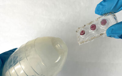
(a) Schematic of the ultrasound modulated tomography setup based on heterodyne parallel speckle detection (HPSD). AOM: acousto-optic modulator, BS: beam splitter, CCD: charge coupled device camera. Two syringe pumps are used for enforcing a controlled blood flow. (b,c) Schematic of the sample set-up in two different experiments. More information here.