After the launch of this article series in March, April was another month of wonderful images from the world of research. This month in pictures takes a look at some of the most dazzling images that were published last month for your scientific and aesthetic pleasure.
It seems April’s theme is particularly black and white. But all is not so colorless, as we see in the first image here:
Intestinal organoids
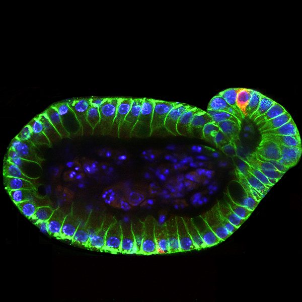
This stunning image featured on the front cover of Advanced Healthcare Materials’ April special issue “Biomaterials in Mechanobiology“, and shows a differentiated organoid with immunofluorescent staining for F-actin (green), nuclei (blue), and paneth cells (red), a marker of intestinal gland, or crypt, formation. Kristi Anseth at the University of Colorado Boulder, who led the study, and her co-workers, believe an understanding of the mechanical dependence of crypt architecture is necessary to instruct homogenous, reproducible organoids for clinical applications.
Filter forests
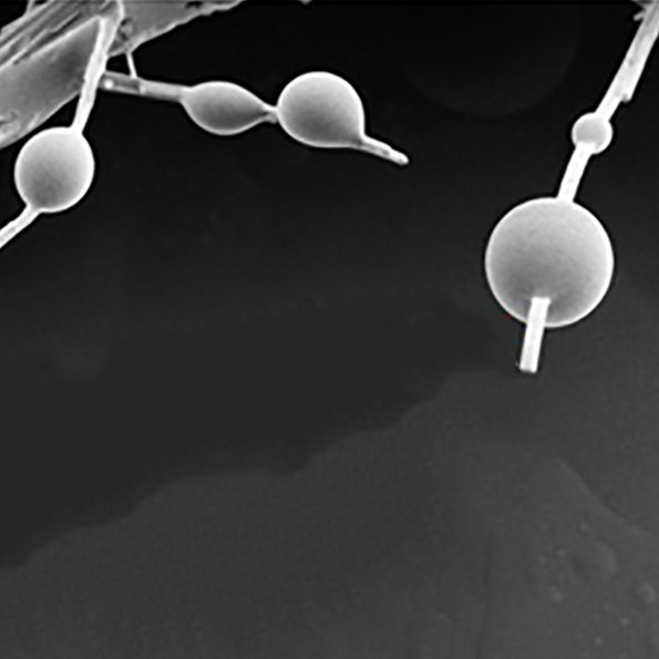
What looks like morning dew on a tree branch are in fact particulate matter from smoke captured on the nanofibers of a renewable air filter material. Swee Ching Tan and co-workers at the National University of Singapore believe a holistic approach is needed for air filtration materials; the innovation here isn’t the ability to filter particulate matter, but to make the filters themselves sustainable and renewable, using less energy-intensive manufacturing processes and raw materials. The image is testament to these eco-friendly materials being as effective as conventional ones.
MOF boxes

What might look good as a minimalist shelving unit wouldn’t be able to store much; these metal-organic framework (MOF) crystals are barely a micrometer across. What they can do, though, is serve as useful precursor materials before annealing with Co@N‐doped carbon composites. Renchao Che and colleagues at Fudan University synthesized these nanomaterials for strong microwave absorption. This work and that of the air filters above can be found in issue 14 of Small.
Imaging a broken heart
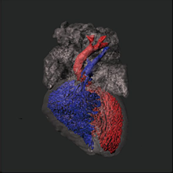
This image is a computer-generated 3D reconstruction based on CT scans of a mouse heart. Yu Nie, Shengshou Hu, and co-workers at the Chinese Academy of Medical Sciences developed computer‐assisted cardiac cavity tracking (CACCT), which can detect the connections between cardiac cavities and identify complicated cardiac malformations in mouse hearts automatically. Blue indicates the right ventricular cavities and pulmonary artery; red indicates the left ventricular cavities and aorta. These were also automatically generated. This work can be found in issue 8 of Advanced Science, and was also featured as the inside front cover of the same issue (not this image).
Micro-mazes
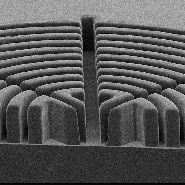
If you’re privileged enough to have an exceptionally large garden, the image above might not be a bad design for your hedge maze. Thankfully, though, some advances in science can be a great social leveler, as this particular maze is only a few hundred micrometers in size. It is the work of Philip LeDuc and colleagues at Carnegie Mellon University, and is obviously not cut from hedges. A technique using polycarbonate heat molding was introduced by them to clone master molds for soft lithography into monolithic, thermoplastic sheets for further soft lithographic fabrication. This technique can be employed to copy a great variety of microfeatures at different scales ranging from the submicron level up to tens of centimeters. The above image is a PDMS microstructure achieved through this very technique, and is featured in issue 16 of Small.