The ability to detect and monitor individual biomolecules has given researchers profound insight into complex intra- and intercellular functions.
Over the past two decades, single-molecule techniques have seen a rapid increase in capability and versatility, which has led to their application within a variety of fields ranging from fundamental biochemistry and molecular biology to genomics.
To consolidate the recent developments in the field, Benjamin Croop, Chenyi Zhang, Youngbin Lim, and co-workers have highlighted the emerging techniques that utilize optical methods for single-molecule detection in a recent WIREs Systems Biology and Medicine Review.
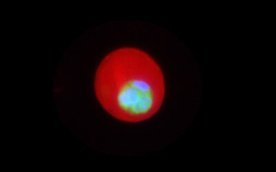
Fluorescent labeling is a popular approach for single-molecule imaging, providing excellent signal-to-noise ratio and the ability to specifically image the biomolecule of interest. New techniques allow researchers to explore the dynamics of biomolecules within cells at faster rates than ever before, providing a clear view of their interactions and behavior.
Other fluorescent imaging methods enable the acquisition of far richer information than ever. This allows scientists to observe rare interactions or mutations, and variations in the populations of biomolecules at the cell-to-cell level.
These techniques have been applied to the detection and potential treatment of various diseases, including neurodegenerative diseases, cancer biomarker screening, and drug development.
As contrasted with fluorescence-based single-molecule experiments, label-free approaches can investigate the unperturbed dynamics of biomolecules. Tether-free techniques enable the observation of a biomolecule without physically binding it to a surface, which can affect its function.
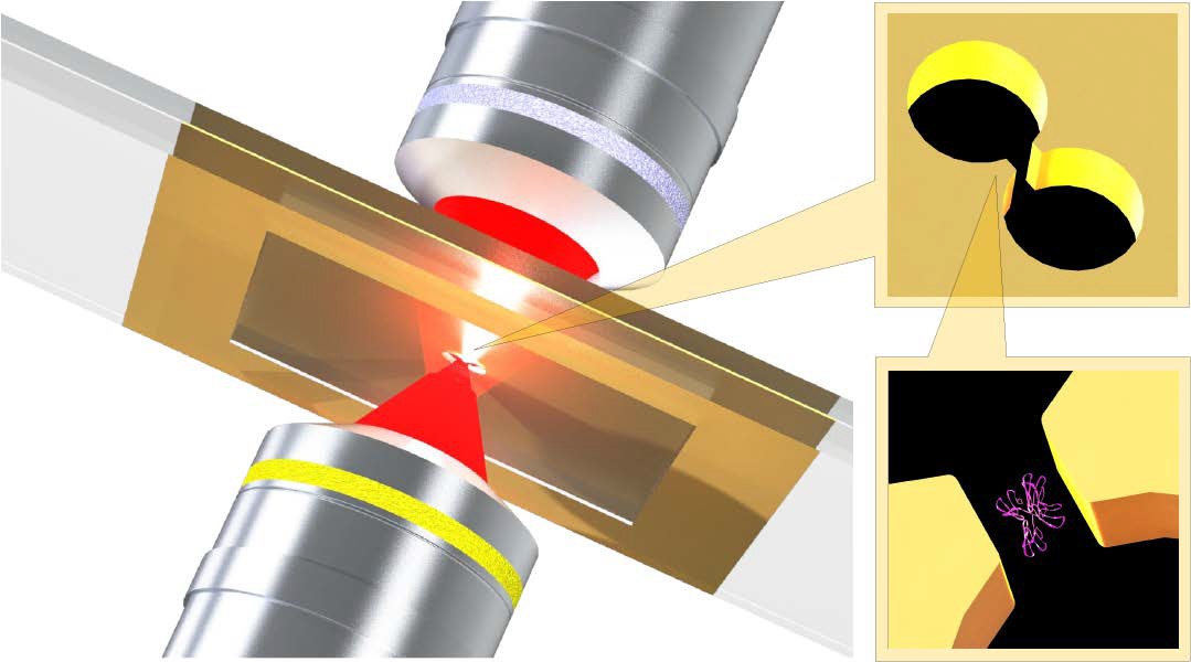
Optical trapping techniques have been improved by the use of nanoapertures, which reduce the risk of damaging the biomolecule and allow researchers to investigate the size, binding behavior, or other dynamics of a freely diffusing biomolecule.
It is possible to detect the small amount of light scattered by individual biomolecules, which enables label-free imaging. The scattered signal is proportional to the mass of the biomolecule, which allows this technique to serve as a nondestructive alternative to mass spectrometry.
Topics in the review include fluorescent and label-free methods for monitoring biomolecular interactions and changes in conformation, as well as methods of protein detection and analysis. To assist researchers in finding a technique suitable for their needs, the strengths and limitations of techniques are presented, as well as potential future application areas.
Kindly contributed by the authors


