
The past decades have seen rapid development of noninvasive methods for skin cancer screening. As skin cancer is the most common type of cancer, there is a broad public interest to improve the diagnostic procedure. In particular, malignant melanoma (MM), the most dangerous type of skin cancer, receive more and more attention as the incidences are increasing worldwide, especially in regions with high ultraviolet (UV) light exposure and population with Caucasian skin type. In addition, excessive sunbathing and use of sunbeds, also lead to unnecessary UV exposure and increase the probability for mutation of melanocytes. MM is hard to treat when diagnosed at a late stage where the pathogen cells are spread in the cardiovascular or lymphatic system. Therefore, early diagnosis and complete surgical excision of the MM are essential for improving of the prognosis of patients in general.
Currently skin cancer screening consists of an initial visual inspection of melanocytic nevi, for example, via a dermatoscope. Typically, for suspicious lesions, a surgical excision is recommended, usually resulting in a large number of false positives. The excision and the past‐excisional histopathological analysis is invasive and painful, time consuming and thus expensive. For all these reasons, there is a strong demand for fast, noninvasive and objective techniques capable of preoperative skin cancer diagnosis, 3‐dimensional (3D) growth and morphology tracking as well as reliable margin assessment of suspicious melanocytic nevi required for the further excision.
A variety of diagnostic modalities has been applied for noninvasive skin cancer detection so far including optical coherence tomography (OCT) and optoacoustics (OA). Another modality is Raman spectroscopy which has the potential to not only distinguish cancerous tumors from the benign lesions, but also for the identification of the different skin cancer types. It appears that one technique alone will not be able to reliably determine the surgical margins for a wide range of melanoma thicknesses noninvasively, and, therefore, a combination of such techniques is a more promising route to realize such a system in future.
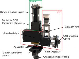
Rendered image of the combined OCT‐Raman probe with an applicator and a spacer ring
A team of researchers from the Leibniz University in Hannover, Germany did just that: they set up a new multimodal system for rapid, noninvasive in vivo skin cancer screening, combining optical coherence tomography (OCT) and optoacoustic (OA) modalities to provide precise tumor depth determination with a Raman spectroscopic modality capable of detecting the lesion type and, thus, providing diagnostic capability. “The three modalities were combined for the first time to our knowledge to perform simultaneous (OCT, Raman) and colocalized (OA) measurements on skin lesions including MM in preclinical trials. The system appears suited as a future medical tool to improve skin cancer screening and early diagnosis, in particular, as all measurements were performed below the admitted maximum permissible exposure levels” according to team member Arthur Varkentin.


