")
Following fertilization in mammals, the embryo develops while passing through the oviduct (fallopian tube) before implanting in the uterus. This period of preimplantation development consists of several critical stages with distinct cellular and molecular activities defining the health and viability of the embryo.
The mouse is a well‐established model with powerful genetic engineering strategies for studying mammalian reproduction and embryonic development. However, our current understanding of the preimplantation developmental process has been obtained primarily from ex vivo culturing experiments; in vivo analysis of preimplantation embryos in the oviduct, their native growing environment, is largely absent.
Unfortunately, there is still a lack of imaging techniques that are able to generate the desired visualizations through tissue layers. The oviduct supplies a complex biochemical and biomechanical environment for the early embryo, which is hard to model under ex vivo settings. This technical hurdle prevents an understanding of the natural process of preimplantation development and limits the study of interactions between embryos and the oviduct. This missing knowledge is significantly valuable for improving the management of infertility and in vitro fertilization (IVF) in humans.
More traditional modalities, such as bright-field microscopy and interference-based quantitative phase microscopy, provide a set of useful tools for live investigations of preimplantation embryos, but removal of embryonic cells from the female reproductive tract is required. In doing so, we lose the potential for in vivo study of preimplantation development in the oviduct.
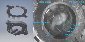
The dorsal intravital window for in vivo optical coherence microscopy (OCM) imaging of the preimplantation embryos. (Left) illustration of the window and protective metal cover. (Right) a close‐up picture of the window taken in vivo.
A group of scientists from the Baylor College of Medicine in Texas have demonstrated the feasibility of using optical coherence microscopy (OCM) to image and stage the mouse preimplantation development in vivo. Using OCM through a dorsal imaging window, characteristic morphological features of oocytes, zygotes, and preimplantation embryos can be well resolved both in vitro and in vivo. The imaging quality of OCM is comparable with the traditional bright‐field microscopy in vitro.
“This study opens new exciting opportunities to understand the early developmental process naturally taking place in the oviduct and to investigate interactions of the oviduct with preimplantation embryos, which may provide insights for better management of infertility and the further improvement of assisted reproductive technology” according to team member Irina V. Larina.


