
Optical coherence tomography (OCT) is an emerging technique for cross‐sectional imaging widely used in a nondestructive investigation of the internal structure of different (first of all biological) samples. Several functional extensions to the technique were proposed which allow extraction from OCT data information in addition to structural cross‐sectional images. Among the set of functional extensions of OCT, one of the most promising are various techniques that allow one to visualize flows inside the tissue. The key factor for distinguishing between flow and static clutter in the tissue is the fact that presence of the flow leads to bigger variability in OCT signal in comparison with a signal from static background. Since the first demonstration of Doppler OCT various modalities searching for variability in OCT signal have been proposed.
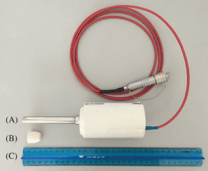
(A) OCT scanning probe prototype used for oral mucosa microvasculature visualization in the present study, (B) the expansion tip for increasing the contact area between the probe and an object for cutaneous microvasculature visualization and (C) the scale
One of the key requirements of all these methods is the ability to eliminate possible bulk tissue displacements, induced by patient’s respiratory and cardiac motions at data acquisition step or compensate them at post-processing step. Although several bulk motion correction approaches were introduced, they all implicitly apply some constraints at the type and magnitude of bulk tissue motion as well as that these methods have poor performance in case of displacements of different magnitude and direction at different depths of an object.
Online visualization of obtained vessels cross sections may be considered as one of the crucial steps in the development of convenient OCT‐based angiography system for everyday clinical use. Such visualization provides feedback to the system operator, allowing one to adjust scanner head in a way that enables vessels to be visible and to search for a proper scanning site, and also provides additional motivation to the patient, who can immediately see the results of the examination.
A Russian research team developed an OCT‐based system for real‐time angiography B‐scan visualization, capable of using with a hand‐held probe, which makes it convenient for everyday clinical use. The key features of the system are the method for compensation nonuniform tissue displacements and algorithm, which computes and visualizes 1 angiographic B‐scan while acquiring data for the following structural OCT B‐scan, with angiography processing along slow axis. According to team member Alexander Moiseev “two major problems were solved during the development: firstly, compensation of specific natural tissue displacements, induced by contact scanning mode and physiological motion of patients and secondly, online visualization of vessel cross‐sections to provide feedback for the system operator”.


