
Recent decades show cardiovascular disease ascending as a major cause of mortality and morbidity worldwide, especially in Western countries. Induced by plaque build-up that narrows the vessel lumen and blocks blood flow to the myocardium, coronary artery lesions may lead to myocardial infarction, which may further progress to heart failure. To understand the atherogenesis that is strongly affected by the stresses and strains in the cells and fibers of the arteries, knowledge of the mechanical properties of coronary arteries is fundamental.
In addition, insights into mechanical responses of fresh arteries to external stimulations can also greatly clarify vascular physiology which can serve as a reference for understanding initiation, progression and clinical treatment of vascular disease. The mechanical properties of artery tissue are determined by its 3 major components: collagen fibers, elastins and vascular smooth muscle cells (VSMCs).
Coherent Raman scattering (CRS) microscopy, based on either coherent anti-Stokes Raman scattering or stimulated Raman scattering (SRS), is an emerging vibrational imaging technique for biomedical tissues in living systems. With significantly improved imaging speed and detection sensitivity, CRS techniques have been recently reported to allow real-time non-invasive imaging of living cells and organisms with chemical selectivity in a label-free manner.
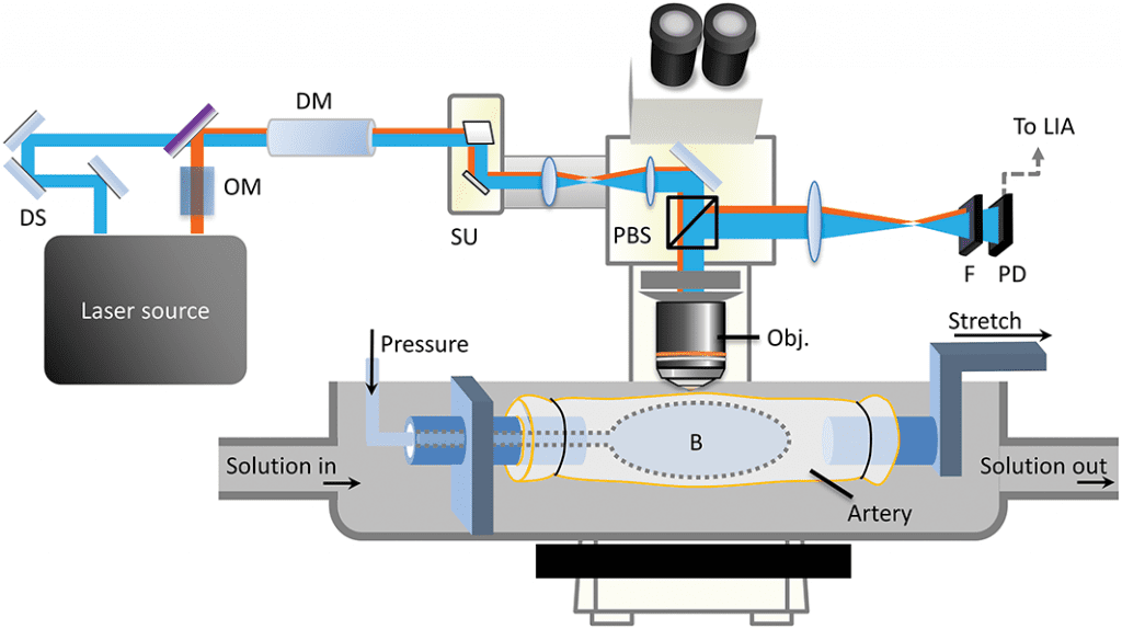
Schematic of the epi-SRS setup for VSMC imaging in fresh coronary artery with controlled loading pressures.
Several researchers from the US teamed up to understand the specific geometries of VSMC microstructure under physiologic state and their responses to external stimulations, as well as the overall mechanical behaviors of vessel wall. To achieve this, the team set up a water bath-based epi-SRS microscopic platform that was designed for simultaneous mechanical loading-functional imaging to quantify the deformation of VSMCs in living coronary arteries.
The morphologic deformation of VSMCs in response to mechanical stress, and the relaxation of VSMCs during vasodilation, were revealed for the first time. “Knowledge on active VSMC deformation under stress is essential for better understanding of VSMC functions in arteries and for development of new strategies for cardiovascular disease treatment” according to team member Ghassan Kassab. The study provides a more accurate framework for the biomechanics of blood vessels, and may open a new door for functional imaging of blood vessel tissues under physiologic and pathologic conditions.


