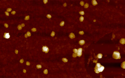
In medical applications including biomedical imaging, drug delivery, and cancer treatment, gold nanoparticles have attracted a great deal of attention. However, the effect of nanoparticles on cellular stress is not fully understood.
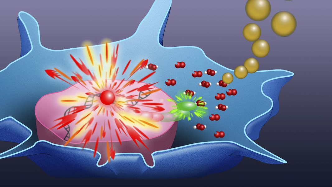
In their article in Small, Prof. Nan Ma from the Institut für Biomaterialforschung and Prof. Ulrike Alexiev and colleagues from the Freie Universität Berlin and the Helmholtz Virtual Institute—Multifunctional Biomaterials for Medicine present a technique that enables highly sensitive detection of the production of intracellular reactive oxygen species (ROS) in cells and tissue.
Prof. Ulrike Alexiev: “In the mitochondria of our cells, enzymes transport electrons along a chain of redox reactions to oxygen for energy conversion in a process called oxidative phosphorylation. As a byproduct, highly reactive molecules are formed, which are called reactive oxygen species (in short ROS).
These reactive oxygen species are on the one hand involved in cellular regulation, but on the other hand related to aging, diseases, and therapeutically adverse effects, because the overproduction of ROS can damage DNA, proteins, and lipids, thereby impairing normal cellular function and potentially leading to cell death. Nanoparticles can also lead to an overproduction of ROS, which is a major concern for nanotoxicity.”
Jens Balke: “We use fluorescence lifetime imaging microscopy (FLIM) to detect ROS-activated dye fluorescence from a ROS reporter dye called CellROX Green—a technique we termed “FLIM-ROX”—for low-level ROS identification. This technique provides an understanding of ROS-associated nanotoxicity of nanoparticles in single cells and tissues, and enables high-throughput analysis.”
Prof. Ulrike Alexiev: “The precise assessment and monitoring of enhanced ROS levels in healthy and atopic dermatitis skin demonstrates the potential of this technique for the quantification of spatial ROS distribution in vivo, for instance, as a consequence of nanoparticle application or as a potential trigger for controlled release of drugs from nanocarriers.”
To find out more about this technique for elucidating ROS-associated nanotoxicity, please visit the Small homepage.


