
Distinguishing tumors from normal brain cells is important in diagnosis and is particularly challenging in glioma (a type of tumor that occurs in the brain and spinal cord) surgery due to the lack of clear interfaces between healthy and affected cells.
Prof. Marie Louise Groot and her team from Vrije Universiteit Amsterdam demonstrated the potential of label‐free third harmonic generation (THG) microscopy in combination with automated image analysis for diagnosing glioma tumors during surgery. The aim was to develop a “snap-reading” pathology diagnostic tool that would provide real-time information to the pathologist on the cancerous nature of an excised brain tissue sample.
THG is a form of nonlinear imaging and has advantages over other spectroscopies, such as Raman spectroscopy, in that it directly visualizes cellular morphology in tissue and is technically much simpler. The authors presented specific THG image characteristics for normal white and grey matter of the brain, as well as for high- and low-grade gliomas. The study resulted in the successful assessment of low-grade (slow growing) and high-grade (fast growing) glioma, which matched closely with assessments made by trained neuropathologists.
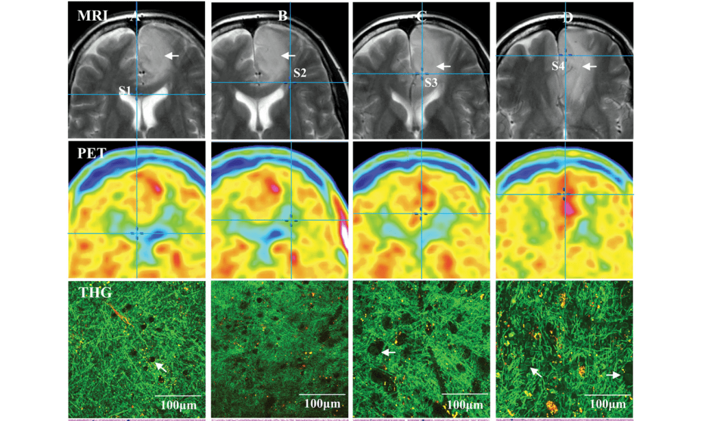
THG imaging of a grade II astrocytoma with corresponding MRI, and PET images.
The condition of cells was validated by quantitative comparison of THG imaging with fluorescence microscopy of nucleus‐stained samples indicating that THG reflects true tissue cellularity, which differentiated normal brain from glioma infiltration, with 96.6% sensitivity and 95.5% specificity, and in good (93%) agreement with the neuropathologists. Four tissue samples taken from one patient were analyzed and there was a positive correlation between cellularity assessments made by THG, preoperative magnetic resonance, and positron emission tomography imaging. All results of this study are well validated with independent data, providing significant statistics and thus demonstrating the relevance of the proposed approach.
Prof. Groot and her co-authors clearly showed that quantitative THG microscopy has the potential to improve the accuracy of brain tumor surgery.


