Brain-machine interfaces are the key to curing many neurological disorders and diseases, as well as the future integration of modern technology with biological systems.
The long-term stability of brain-machine interfaces is a major challenge due to the huge mechanical mismatch between soft neural tissues and hard electronics. Recent progress in the field of soft and stretchable electronics has shown that metallic nanomaterials in combination with rubbers facilitate the fabrication of soft electric conductors with similar mechanical properties as living tissue.
However, current limitations in materials and fabrication processes have prevented soft electronics from reaching their full potential in brain-machine interface applications.
In their paper published in Advanced Materials, Klas Tybrandt (Linköping University, Sweden and ETH Zurich, Switzerland), Dion Khodagholy (Columbia University and New York University, USA) and their colleagues demonstrate soft electrode grids for the long-term chronic recording of neural signals. The high-density grids can resolve high spatiotemporal signals during three months of implantation with preserved signal quality.
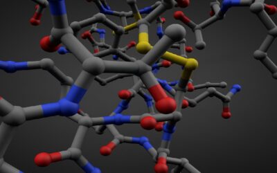
The grids are based on a new composite material that consists of gold-coated titanium dioxide nanowires embedded in a silicone elastomer. The composite combines high conductivity, excellent electromechanical properties, long-term stability, and biocompatibility, making it ideal for biomedical applications. The composite is processed into high-density electrode grids by a novel fabrication method, which is designed to preserve the functionality and biocompatibility of the composite material.
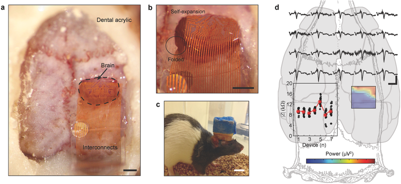
Implantation and in vivo neural recording of soft SEGs. a) Intraoperative microphotograph of soft SEG showing the probe in a craniotomy lying on the surface of the brain. Note the hydrophobic nature of the device demonstrated by a drop of water on the ribbon area. b) Zoomed-in microscopy image showing the ability of the probe to be inserted in smaller craniotomy than the probe geometry. c) Freely moving rat with chronically implanted SEG. d) Intraoperative recording of SSEPs using the soft SEG.
The chronically implanted electrode grids show excellent performance due to their conformal contact with the tissue.
The deformability also allows for insertion of the grids through small openings, followed by self-expansion of the grids.
Klas Tybrandt believes that the combination of the novel material and fabrication process opens up for many new applications stating, “Soft electronics are attractive for all kinds of electronic interfacing of tissue. The properties of our developed material make it ideal for a wide range of applications towards the central and peripheral nervous system”.
Kindly contributed by the authors.