
Many diseases affecting neurological functions can be linked to pathological alterations of the cerebral vasculature. Some of the alterations are known to cause the disease, support and accelerate its spread, while in many other cases, the interaction between the disease etiology and vascular pathology is only poorly understood. This critical link between vascular changes and disease is increasingly targeted by preclinical studies in mouse models mimicking the pathological changes in human cerebrovasculature.
Imaging techniques and various intravital techniques are commonly used to provide metabolic and functional brain images in humans, but have several limitations like necessitating exogenous contrast agents. Optoacoustic (OA) imaging techniques have recently provided unprecedented insights into the deep‐tissue anatomy and physiology of animal model organisms due to the unique combination of rich and endogenous optical absorption contrast and weak scattering of US waves in biological tissues.
Imaging performance yet greatly differs among the different OA microscopy systems which all have different challenges. To address these unmet challenges, an international team of researchers of the Institute of Biological and Medical Imaging in Germany developed a fast functional OA microscopy method that uses a Pockels cell (PC)‐based wavelength switching for rapid acquisition of the spectral data.
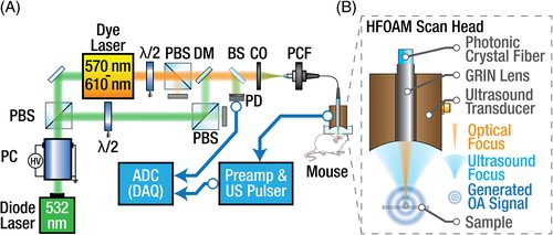
Schematic diagram of the dual‐wavelength hybrid biomicroscopy system.
This novel hybrid OA and functional ulstrasound biomicroscopy approach capable of large‐scale morphological and functional brain imaging was realized using dual‐wavelength illumination. Rapid wavelength switching in combination with fast mechanical scanning and a flexible, highly sensitive scan head design have enabled the high‐speed visualization of both morphology and oxygenation status of large‐scale vascular networks with lateral resolution of 12 μm across FOVs covering the entire murine cortex. The flexible scan head design allows for concurrent high‐resolution pulse‐echo US scans, providing additional anatomical information.
“In conclusion, the presented biomicroscope can bridge the gap between localized microscopic observations and lower resolution macroscopic imaging in whole mouse brains, thus offering a new scalable multimodal tool for investigating healthy and pathological neurovasculature.”, according to team member Daniel Razanksy.


