Scientists have developed a new technique that will allow them to create small and flexible vascular grafts for coronary artery bypass procedures, making the treatment of coronary heart disease safer.
The mortality rate associated with heart disease has surged since 2019, with the World Heart Federation estimating a 60% rise in deaths associated with cardiovascular disease. This makes the need for efficient and effective medical interventions a pressing concern.
Treatments for cardiovascular disease may range from medications to help manage the condition to lifestyle changes, such as an improved diet and more exercise. However, in extreme cases, surgical intervention becomes necessary.
Grafts provide life-saving interventions
One of the most common forms of cardiovascular disease is coronary heart disease, caused when arteries become clogged, potentially leading to heart attacks, stroke, and heart failure. The most commonly performed surgical procedure in these cases is coronary artery bypass, with 400,000 such procedures taking place each year in the United States alone.
Coronary artery bypass involves taking a blood vessel from another part of the body, commonly the leg or arm, then attaching it to the coronary artery above and below the blockage. This redirects the flow of blood, thus allowing it to bypass the blockage. These new blood vessels are called “grafts,” and they help to restore the blood supply to the cardiac tissues, which face a shortage of oxygen and nutrient supply as a result of blocked coronary arteries.
However, when grafts created using human tissues are not viable, surgeons can turn to artificial or synthetic versions. Currently, marketed synthetic grafts are made of materials called “perfluorinated and polyfluorinated alkyls” (PFAs).
These cause the patient to have an immune response to the graft, and bans are currently planned to remove around 10,000 PFA substances from the market due to issues with environmental contamination, requiring a new, safe graft alternative.
A safer, more flexible graft
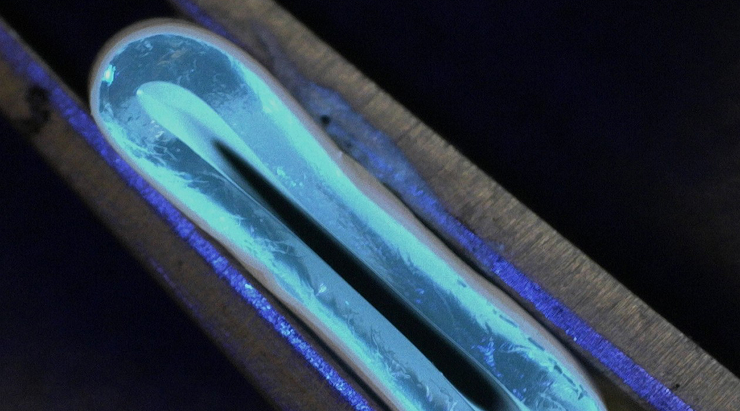
The team’s new graft, suggested in a paper published in Advanced Materials Technologies, uses a combination of extrusion printing and electrospinning to deposit nanofibers over a printed hydrogel tubular body. This provides a synthetic graft with tensile strength and flexibility comparable to natural human arteries.
“The newly developed extrusion printing system uses a rotating vertical mandrel [a tapered tool used to form rings] and an extrusion tip to print hydrogel, which includes mostly water,” said the study’s author and University of Edinburgh School of Engineering researcher Faraz Fazal. The printing parameters were carefully controlled to print a tubular construct around the rotating mandrel.
“Subsequently, the grafts were reinforced by using the electrospinning technique, in which nanofibers 200 times thinner than human hair were deposited over the printed hydrogel,” he said.
Fazal added that the flexibility of the vascular grafts made from this new method can be easily altered by changing the composition of the nanofibers used to reinforce them. That means they are more compliant and less likely to lead to rejection.
“It is challenging to produce small-diameter vascular grafts [grafts with diameters less than 6 millimeters]. Our method offers a way. We can also produce grafts down to 1 mm, and we can also gradually increase or decrease the diameter, so it can truly mimic the blood vessels in our body,” said corresponding author Norbert Radacsi, also from the University of Edinburgh. “Further, we can use induced pluripotent stem cells [“immature” cells capable of giving rise to several different cell types] from the recipient’s body to reduce the immune response and possible rejection.”
Radacsi added that the fact the method does not require any PFAS makes it safer than current interventions and means it is unaffected by the planned ban. However, before making it into the surgical room, the electrospun nanomaterial coated hydrogel graft still needs more testing before it can be brought to market,.
“We had to optimize our printing technique, which took us about a year. Then, we did the tensile testing, burst pressure measurement, compliance, wettability, microstructure, and cell biocompatibility testing on the products, both with and without the nanofiber layer,” Radacsi explained. “We now need to conduct animal tests to evaluate the safety of our vascular grafts, followed by human trials. We are currently preparing a proposal for using our grafts in pigs.”
A future beyond just treating the heart
With the testing that has taken place thus far, the team has been pleasantly surprised by many aspects of the system’s design and performance.
“We were surprised how well the printed layers fused and gave us an optically transparent product before the nanofiber coating. We were also surprised how much the burst pressure and compliance improved when the nanofiber reinforcement was used, reaching similar numbers to native human arteries and veins,” Radacsi said. “Finally, it was unexpected that even though we built the vascular grafts horizontally, the failure upon bursting it was always a vertical line.”
The team’s new graft and its creation technique could have implications for medicine beyond the surgical treatment of coronary heart disease, too.
“Since every organ needs blood vessels to supply oxygen and nutrients, our small-diameter vascular grafts will help with printing large organs, even ones with complex vasculature,” Fazal concluded. “This is crucial for 3D printing organs, like heart, kidney, liver, etc.”
Reference: Norbert Radacsi, et al., Fabrication of a Compliant Vascular Graft Using Extrusion Printing and Electrospinning Technique, Advanced Materials Technologies (2024). DOI: 10.1002/admt.202400224


