Three different types of pigment cells, arranged in superimposed layers, are responsible for skin pattern formation in adult zebrafish: black melanophores, yellow xanthophores and silvery iridophores. Melanophores are present in the bottom layer, iridiophores in the middle layer, and xanthophores are found in the top layer. Melanophores are only present in the dark stripes whereas both xanthophores and iridophores – in different shapes and densities – are located throughout the entire body.
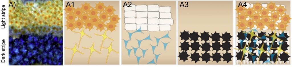
Organization of pigment cells in the trunk skin of adult zebrafish. A: Close-up view of adult skin. A1-A4: schematic representation of xanthophores (A1), iridophores (A2), melanophores (A3), merged view (A4).
In their review in BioEssays, Nüsslein-Volhard and Singh discuss findings from clonal analyses to reveal the developmental origin and fate of these different pigment cells: They all originate from neural crest derived stem cells. At the larval stage, zebrafish are almost completely transparent. The characteristic pigmentation pattern only begins to appear during metamorphosis – a phase that starts about three weeks post fertilization and lasts for about one month. During this period, adult pigment cells appear and start to cover the fish. Surprisingly, pigment cell progenitors remain multipotent at least until this stage.
However, despite their shared lineage, each pigment cell type behaves differently. Iridophores and xanthophores are capable of proliferating and spreading in the skin as differentiated pigmented cells, whereas pigmented melanophores hardly ever divide or migrate. Interestingly, it was also found that contact-dependent interactions between different pigment cell types – rather than long-range molecular gradients – are responsible for the observed self-organization of the color pattern. These direct cell-cell interactions seem to be mediated by potassium channels as well as gap junctions, but the underlying molecular mechanisms still remain to be elucidated.