Mammary organoid. Image credit: Shuichi Takayama et al.
While science is of course a logical and technical discipline, a great deal of creativity is required to make scientific breakthroughs. Problem solving, thinking outside the box, pushing boundaries, and exploring new horizons are descriptors that could just as easily be used for science as they could art. Though they are thought of as separate, combined it’s clear the impact one has on the other. This month our editors have selected some of the most striking images from our journals that capture this creative and innovative spirit.
Detecting gamma rays with giant perovskite crystals
Gamma rays are the highest energy members of the electromagnetic spectrum and when they come into contact with biological materials, the can do a whole lot of damage. Detecting them in settings like nuclear power plants or medical facilities is therefore extremely important. In Advanced Science, Pavao Andričević, László Forró, and their co-workers have developed a technique for growing a massive crystal (seen here) made from a material class called a “perovskites” that act, in this case, as an effective detector that is able to heal any defects, allowing it to operate for a long period of time without degradation.
Artificial nerve grafts never looked so good
Peripheral nerve injuries are difficult to heal, but Joaquim Oliveira from the University of Minho and co-workers have devised an alternative strategy to solve some of the problems related to biological grafts. In a study published in Advanced Healthcare Materials, artificial nerve guidance conduits were made of silk fibroin that can incorporate growth factors and promote effective regeneration after injury. This stunning image was chosen as the cover of the journal’s January special issue.
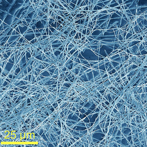
Yarn muscles
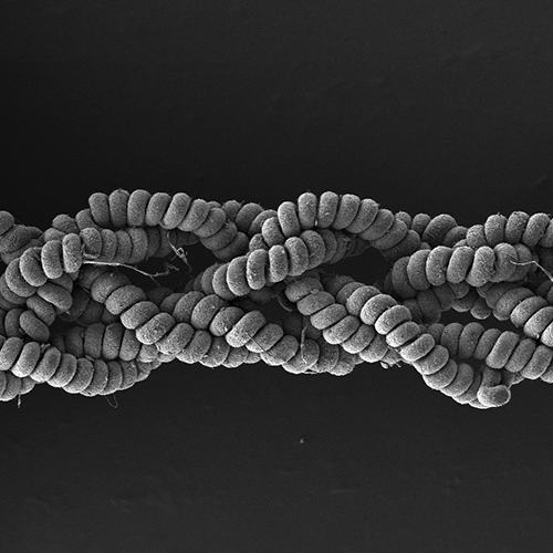
Yarn that behaves like biological muscles is not as far-fetched as one might initially think. This SEM image, published in Small, shows a braided artificial muscle yarn created by Jiangtao Di, Qingwen Li, and their collaborators that combines the advantages of ionic liquids and nanofiber polymers. They’ve even shown their muscle’s capabilities with a muscle building demonstration (complete with tiny dumbbell!) that we thought was only fitting to share.
Growing new tissue
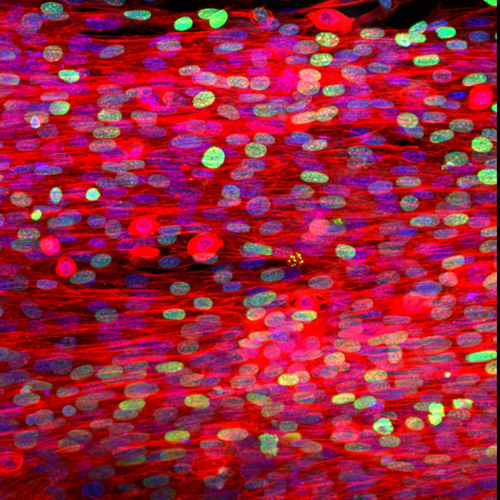
Skeletal muscle has an inherent capacity for spontaneous regeneration. However, recovery after severe injuries is limited. In Advanced Materials, Polina Anikeeva, Seung‐Woo Cho, and team report a new tissue regeneration strategy that employs direct cell reprogramming in combination with a new hybrid scaffold. This image is part of a series of experiments showing the enhanced myogenesis of induced skeletal myogenic progenitor cells (iMPCs) when using a muscle extracellular matrix (MEM)–polycaprolactone (PCL) fiber‐based hybrid construct.
Moons or marbles?
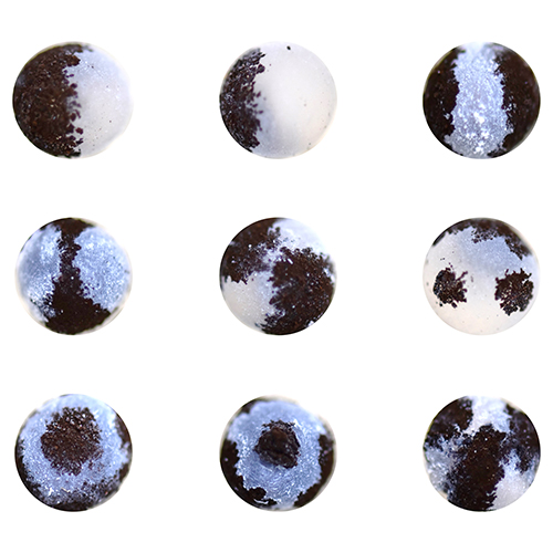
The answer is actually liquid marbles, which are droplets that have been wrapped in solid particles for applications in fluidics and soft device applications. In a study published in Advanced Functional Materials, Masanobu Naito, Mizuki Tenjimbayashi, and their co-workers report a general strategy for the flexible patterning of functional particles on droplet surfaces in a patchwork‐like design, as seen in the above image.
Flexible films
In Advanced Materials, Alexander Balandin and co-workers demonstrate that quasi‐1D van der Waals materials like TaSe3 (shown in the above SEM image) can be used as metallic fillers in polymer composites, providing excellent electromagnetic shielding capabilities. These films are flexible, light‐weight, corrosion resistive, electrically insulating, and inexpensive, and will be important to current 5G and future communication technologies.
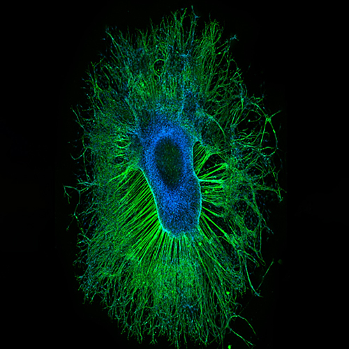
Organoids monitor cancer invasion
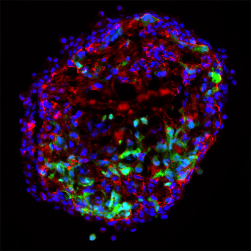
This breathtaking image by Shuichi Takayama and colleagues captures a mammary organoid built to act as an in vitro model for studying “cancer invasion” of cells. In the study published in Advanced Healthcare Materials, the researchers seeded cancer cells on the outside of preformed organoids and their invasion was monitored. The hope is that these high throughput platforms will open new opportunities for convenient, standardized, and physiological cancer cell invasion studies. This image was selected for the journal’s February issue, which is dedicated to Professor George Whitesides.
Silica glass microstructures
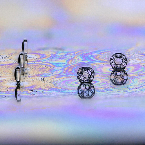
Fused silica glass is the material of choice for many high‐performance components in optics, especially fused silica microstructures for microoptical and biomedical applications. In a study published in Advanced Materials, Frederik Kotz and his colleagues report true 3D structuring of transparent fused silica glass using direct laser writing, achieving structures with micrometer resolution. Above we can see three fused silica upright microlenses (on the left) directly printed on a silicon substrate with three Wigner–Seitz cells printed in IP‐Q; the structures are roughly 400 µm.
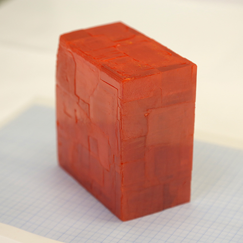
Single crystal X-ray detectors for better imaging
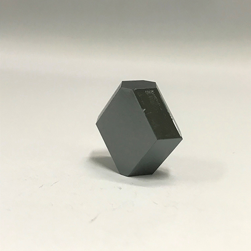
In a recent study published in Advanced Materials, a team led by Yucheng Liu, Mercouri Kanatzidis, and Shengzhong (Frank) Liu report an inch-sized, high-quality triple cation and mixed halide perovskite single crystal for X-ray detection. They demonstrate their crystal’s superior capabilities, which will allow clinicians to obtain faster, higher resolution X-ray imaging for diagnostics.
To see more articles in this series “This month in pictures”, click here