A new finding challenges the classical understanding of the brain’s protective outer layers. The meninges, as they are known, include three shock-absorbing membranes that rest between the brain and skull. Now, researchers report finding a fourth meningeal layer that they have termed the subarachnoid lymphatic-like membrane (SLYM).
“This is a very interesting and thought-provoking work, proposing that meningeal layers are not as simple as we thought they are and I think this work may just be the tip of the iceberg,” said Jonathan Kipnis, a researcher at the Washington University in St. Louis, in the United States, who was not involved in the study.
Why it took so long to find
The three known meningeal layers are termed the dura, arachnoid, and pia maters, ranging from superficial to deep. The newly discovered layer was found to lie between the arachnoid and pia maters, dividing the subarachnoid space. This area between the arachnoid and pia maters, called the subarachnoid space, contains connective tissue, blood vessels, and large spaces filled with cerebrospinal fluid (CSF). This colorless fluid flows within and around the brain and spinal cord, bringing in nutrients and filtering out waste.
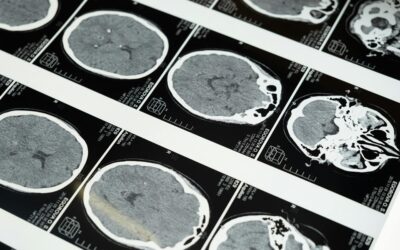
Thus far, the SLYM’s imperceptible width — running as little as one cell thick in some parts — had escaped the notice of older imaging and experimental techniques. Moreover, the faint layer dissolved when the brain was detached from the skull during a post-mortem examination and was undetectable with clinical brain imaging used in the living.
Maiken Nedergaard, a researcher at the University of Rochester Medical Center in the United States, and colleagues first visualized the previously unknown layer in mice using green fluorescent dyes that helped light up the membrane using an advanced microscopy technique. While most experiments were conducted in mice, the researchers also observed this protective layer around the adult human brain.
What does it do?
On testing if this membrane was permeable, the researchers found that only very small molecules were able to pass through. This meant the transport of large peptides and proteins by CSF through the membrane and across the subarachnoid space appeared to be size restricted by the SLYM. Further, when the SLYM was damaged due to a traumatic brain injury, it became leaky, losing its ability to restrict the passage of large molecules.
Researchers suggest that the membrane likely helps separate waste from the CSF. Small proteins like beta-amyloid and tau, whose aggregation is implicated in Alzheimer’s, might be cleansed out of the brain with the help of the SLYM. Any damage to the membrane possibly limits the segregation of these small peptides, contributing to the development of disease, the researchers theorize.
Next, the researchers analyzed if the SLYM was separate from the other meninges. They found that the SLYM was similar in form and function to a kind of layer called the mesothelium. Such membranes surround organs such as the kidneys in mice and the lungs and heart in humans. Mesothelia are thought to reduce friction between tissues, especially in organs that are susceptible to constant movement.
Besides cushioning and lubricating organs, mesothelia often serve an immune function. The SLYM, packed full of immune cells, is seemingly not very different from other mesothelia.
“I think this study makes us keep open minds when studying meningeal layers,” added Kipnis. “This part of the brain/cranium has not been as studied, but this area is important for immune surveillance of the brain, and I am excited that more and more labs are drawn to study meninges these days.”
SLYM and immunity
The SLYM appeared to contain large numbers of myeloid cells, including white blood cells and macrophages. As a result, the SLYM may be well-positioned to have an immune monitoring role. When the SLYM was subjected to inflammation and aging, the types and numbers of immune cells increased, suggesting that it may keep a check on the cerebrospinal fluid.
The researchers speculate that a ruptured SLYM would allow the entry of immune cells from outside the brain to come in contact with it. They suggest that traumatic brain injury may promote inflammation by drawing external immune cells and disturbing the flow of CSF around the brain.
“The discovery of a new anatomic structure that segregates and helps control the flow of cerebrospinal fluid in and around the brain now provides us much greater appreciation of the sophisticated role that CSF plays not only in transporting and removing waste from the brain, but also in supporting its immune defenses,” said Nedergaard in a press release.
Reference: Kjeld Møllgård, et al., A mesothelium divides the subarachnoid space into functional compartments, Science (2023). DOI: 10.1126/science.adc8810
Feature image credit: Dmitriy Gutarev on Unsplash