
 Label-free, non-invasive, noncontact – but high-resolution: Holographic imaging reveals details about morphology, motility, and the concentration of sperm in semen analysis.
Naples (Italy) – Semen analysis is an important diagnostic tool for assessing male reproductive health in both humans and animals. In humans it is mainly used for couple’s infertility exploration or to confirm the success of male sterilization procedures. Animal semen analysis is commonly used in stud farming and farm animal breeding. As abnormal sperm features are the most common indicators for male infertility, there is growing interest in understanding both the spermatozoa morphological alterations and the kinematics/dynamics of the swimming spermatozoa.
For a conclusive analysis of the morphology, motility, and the concentration of the sperm non-destructive methods are needed which neither influence sperm vitality nor have other adverse effects. In addition, the results have to be independent of the experience of the technician and the environmental conditions. In this context, digital holography has shown to be an attractive candidate: it is a label-free, non-invasive, noncontact method and provides high-resolution, enabling the characterization of live specimen. In a review article, scientists from the National Research Council, Naples, and the National Research Council, Pozzuoli, Italy provide an overview on the state-of-art digital holography for semen analysis, present recent achievements and discuss possible new applications in this field.
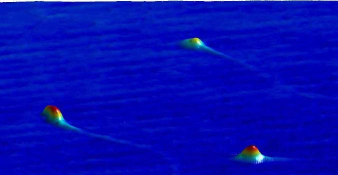
Digital holography is based on the recording and numerical reconstruction of the phase and amplitude of the specimen’s optical wavefront. Thus, 3-D quantitative sample imaging can be automatically produced by numerical refocusing of a 2-D image at different object planes without mechanical realigning the optical imaging system. Consequently, a volumetric field can be reconstructed by means of a single image (the hologram).
3D tracking of the spatial motion of live spermatozoa is possible in order to individuate the right movements of normal spermatozoa. Adding the third dimension will provide a better statistic analysis compared to 2D methods. This is useful both to relate the sperm anomalies with male infertility and to enable differentiation of the healthier spermatozoa. Three-dimensional information on both the morphology and motility of sperm can be obtained.
As simple and cost-effective methods to convert existing microscopes into holographic imaging devices have been recently developed, the method could experience an upswing in the near future.
(Text contributed by K. Maedefessel-Herrmann)
See the original publication: Giuseppe Di Caprio, Maria Antonietta Ferrara, Lisa Miccio, Francesco Merola, Pasquale Memmolo, Pietro Ferraro, and Giuseppe Coppola, Holographic imaging of unlabelled sperm cells for semen analysis: a review, J. Biophotonics 7:10, 779-789 (2015)


