Raman spectroscopy of the retina coupled with multivariate analysis might become a promising tool to study retina diseases in vivo such as retinal tissue damage during inflammation in multiple sclerosis.
 Diseases such as glaucoma and multiple sclerosis damage the retina. Retinal inflammation is primarily mediated by microglia activation, including the release of pro-inflammatory cytokines and the creation of oxidative stress that impairs neuronal function and promotes axonal damage. A more profound knowledge of the molecular changes at the retinal level would be useful to increase the understanding of neuronal loss and axonal transection, and would aid the development of new neuroprotective strategies for brain and retina diseases.
Raman spectroscopy appears to be an attractive candidate for this kind of analysis as it shows the maximum specificity among all optical techniques for detecting molecular changes. Yet the interpretation of Raman spectra is very complex: Biomolecules often have closely related structures and their Raman spectra therefore share groups of bands, making it difficult to deconvolute the contributions of pure molecular components from Raman spectra. Efforts using several statistical calculation methods could not extract meaningful information that would enable characterization of the Raman spectra of the pure molecules in a sample.
Scientists from ICFO – The Institute of Photonic Sciences and the Hospital Clinic of Barcelona, Spain, propose the application of multivariate curve resolution (MCR) to deconvolute pure molecular components from the Raman spectra. MCR represents a novel methodology with promising applicability to study and monitor the biochemical behavior of diseases in vivo. The Spanish researchers applied the method to monitor the molecular profile of the Ganglion cell layer (GCL) over the retinal inflammation process.
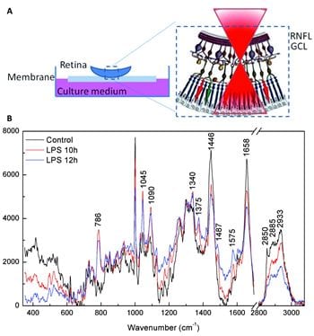
The researchers used an in vitro model of neuroinflammation based on murine retinal organotypic cultures, which preserve cellular composition and tissue architecture while allowing direct in vivo imaging analysis.
First, they performed a Partial Least Squares-Discriminant analysis (PLS-DA) analysis to discriminate between healthy and inflamed tissue at different stages in the inflammation process. Then, they used MCR to deconvolute pure molecular components from the set of experimental Raman spectra that changed during retina inflammation. The scientists monitored the evolution of their concentrations in the tissue over a 24-hour period process.
Six molecular components suffering dynamic changes along inflammatory process were deconvoluted, including the increase of immune mediators (Lipoxygenase, iNOS and TNFa), changes in molecules involved in energy production (Cytochrome C, phenylalanine and NADH/NAD+) and decrease of Phosphatidylcholine.
The authors conclude that Raman spectroscopy combined with multivariate analysis might become a promising application to study retina diseases in vivo.


