
 Researchers from China have modified a nanoprobe with an Iridium complex to sensitively monitor oxygen concentrations in cancer cells. With their method, interfering autofluorescence of biomolecules during measurements is minimized. This is due to special imaging techniques, which distinguish between the short-lasting autofluorescence and long-lasting downconversion phosphorescence of the Ir complex.
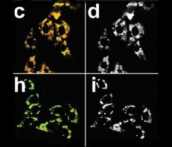
Low oxygen levels in cells (hypoxia) are typically occurring in diseases including solid tumors, inflammatory diseases, and cardiac ischemia. Thus, visualizing hypoxia is an important tool for diagnosis and therapy. The fluorescence intensity and lifetime of the applied core–shell nanoprobes, which have fluorescent Ir complexes in their outer shell, are altered by oxygen molecules. Especially the increase in fluorescence lifetime, from 0.8 µs to 4.0 µs when oxygen is excluded, is significant. Therefore, time-resolved luminescence imaging techniques are able to selectively record hypoxia.
The nanoparticles are taken up by various cancer cell lines and show low cytotoxicity. Additionally, the reported method also allows monitoring oxygen concentration gradients, which is very important for the application in tissues with nonuniform oxygen distribution. Further studies are concentrated on near-IR-excitable nanoprobes for the detection hypoxia in small animals.
Advanced Science is a new journal from the team behind Advanced Materials, Advanced Functional Materials, and Small. The journal is fully Open Access and is free to read now at www.advancedscience.com.


