Engineering a mini-brain is no small feat. Neurons are notoriously delicate, making it challenging for scientists to study the physical properties of brain tissue in the lab without damaging the tissue. But a new paper in Advanced Science reveals the trick to growing brain-like tissue that bends without breaking. The trick? Magnets.
The mechanical signature of the brain’s physical properties, like stiffness, softness, and surface tension, shape how brain cells grow, mature, and form connections with each other. Our brains comprise a roughly half-and-half mixture of neurons and glia, which are cells that provide physical and chemical support for neurons.
Growing truly brain-like neural tissues in a lab requires capturing the realistic “squishiness” and cohesion between cells, but exactly how each cell type drives the brain’s mechanical properties has been a mystery.
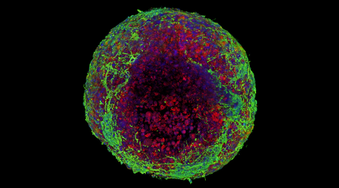
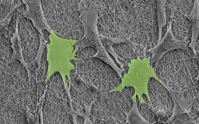
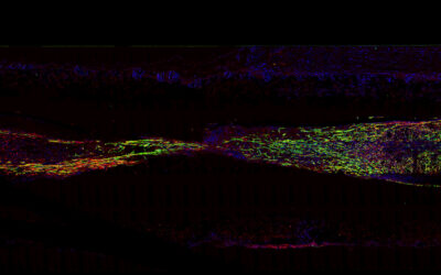
Using tiny magnets to mold a slurry of brain cells into spheres the size of a pencil tip, researchers at the Physio-Chemistry Curie Laboratory in Paris watched as the cells self-assembled overnight. Again and again, neurons migrated to the sphere’s periphery while glia clustered in the middle, forming a core to hold the entire mass together.
According to Jose Efrain Perez, a postdoctoral researcher and lead author of the study, this confirms something neuroscientists have long suspected but haven’t been able to test directly: glia (named after the Greek word for “glue”) are “the binding power of the tissue”, contributing more to brain tissue cohesion and stiffness than neurons.
Molds and magnets
To determine how neurons and glia each contribute to brain tissue cohesion and stiffness, the team first had to successfully mold the cells into a 3D tissue model. “[This] was a challenge in itself,” Perez said. Calculating a tissue’s elasticity and surface tension requires deforming it enough to test its limits without damaging the extremely delicate brain cells.
By immersing tiny steel spheres in hot, liquified agarose gel and letting it cool, the team formed the tiny sphere molds. Then, they carefully pipetted magnetized brain cells derived from mouse embryos into a dish holding the molds.
Using magnets, they lured the brain cells to the molds-like using a magnetic wand to make a Wooly Willy beard out of iron fillings. The cells grow and mature overnight, forming a sphere of brain-like tissue that can be prodded to test its physical properties.
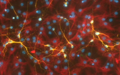
Perez was surprised by how healthy and mature the spheroids were the morning after preparing the cell molds. “We were not expecting to have such beautiful neuronal growth and branching, especially after such a small period of growth,” he said. He suspects that the iron nanoparticles used to magnetize the cells may have helped them establish contact with one another.
To test the relative contributions of neurons and glia to the stiffness and surface tension of brain tissue, they made spheroids with different ratios of each cell type and deformed them under a magnetic field. The tissue’s surface tension increased as the proportion of glial cells increased, confirming that glia are crucial for making our brain’s stick together.
A future in tissue engineering
Despite only growing for one day, the spheroids self-organized into a remarkably stereotyped way, with glia consistently forming a central mass for neurons to gather around.
“It’s fascinating that such a simple model of these two cell types already displays this type of organization,” Perez said. This suggests that the surface tension of the tissue itself may contribute more than neuroscientists suspected to the organization of cells during early brain development.
Moving forward, these simple magnetic brain-like spheroids could be used to ask deeper questions about the biological signals cueing neurons and glia to organize the way they do.
Perez believes that this magnetic engineering technique can be used to build more complex 3D brain models, and even models of completely different tissues, like heart muscle.
“It is a powerful tissue engineering tool,” he said, with a range of promising engineering applications from artificial tissue development to drug discovery.
Reference: J. E. Perez, et al., Surface Tension and Neuronal Sorting in Magnetically Engineered Brain-Like Tissue, Advanced Science (2023). DOI: 10.1002/advs.202302411

