
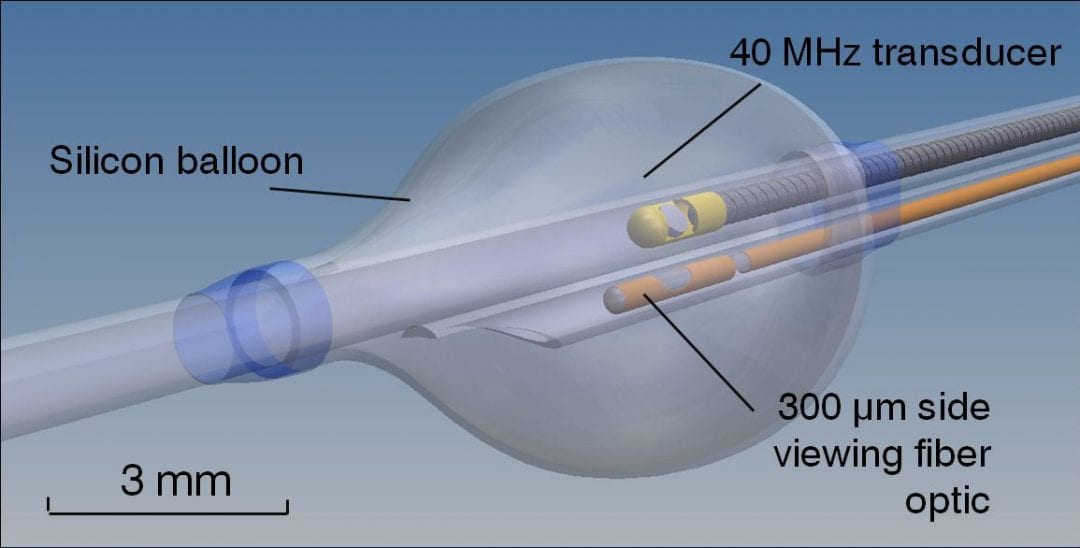
Detail view of the catheter imaging section.
A novel multispectral fluorescence lifetime imaging system with ultrasound guidance could improve intravascular diagnostics and analysis of atherosclerotic lesions. Co-registered FLIm and IVUS data allow 3D visualization of both biochemical and morphological vessel properties.
Coronary artery disease is the leading cause of death in western societies. Although many diagnostic tools have been developed to help predict which patients will suffer cardiovascular complications, current diagnostic techniques have significant limitations. New techniques, such as time-resolved fluorescence spectroscopy (TRFS) and fluorescence lifetime imaging microscopy have demonstrated ability to assess a plurality of diagnostic features associated with arterial wall pathologies including atherosclerotic plaques at risk of rupture. Yet, for clinical practice in intravascular applications TRFS systems and catheters have to be developed that are capable of rapid acquisition of robust time-resolved fluorescence data in vivo from the luminal surface of the artery in the presence of pulsatile blood flow.
To meet this challenge, a novel multimodal catheter system integrating a 40 MHz commercial IVUS and fluorescence lifetime imaging (FLIm) has been developed at the University of California (Davis, USA). Co-registered FLIm and IVUS data allow 3D visualization of both biochemical and morphological vessel properties.
The system uses a fiberoptic for fluorescence excitation-collection. By rotation and pull-back, a fast continuous helical motion scanning (400 rpm, 0.75 mm/s) of TRFS data from luminal surfaces in multiple spectral bands enables fluorescence lifetime imaging (FLIm) of the luminal surface. The team led by Laura Marcu now demonstrated the clinical compatibility of their new diagnostic system by testing it in swine. Their study is the first in vivo intravascular application of a fluorescence lifetime imaging technique.
The researchers conducted these experiments in conjunction with intravascular ultrasound (IVUS) which is the most commonly used intravascular imaging technique in patients. In addition to guiding the TRFS measurement, IVUS provides complementary information on arterial morphology and anatomy. The intravascular rotational catheter combining FLIm with IVUS detection can provide a simultaneous evaluation of the biochemical and morphological features of the arterial vessels.
In 8 seconds, 0.6 cm of arterial segment length could be examined and the autofluorescence features of arterial wall consistently resolved. As expected, the measured arterial wall fluorescence lifetime (~5 ns) was similar to values previously reported for healthy pig arteries when using similar methods for analysis of fluorescence decay. The IVUS played an important role in identifying the arterial regions of interest and in real-time measurement of vessel diameter. Moreover, the IVUS and FLIm data can be co-registered to provide straightforward bimodal image display.


