Real time monitoring of transplanted stem cells is imperative for successful stem-cell based therapeutic regimes.
Advancing this field, Ngen and co-workers develop MRI-based detection of transplanted stem cells that are exogenously labeled with two contrasting agents – high-molecular-weight superparamagnetic iron oxide nanoparticles (SPIONs, T2/T2 * contrast agents, with low diffusion coefficients) and low-molecular weight gadolinium chelates (T1 contrast agents, with high diffusion coefficients).
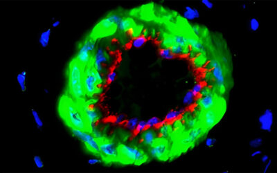
Differences in the diffusion coefficient of the two imaging agents are exploited to produce contrasting MRIs of live and dead cells in vivo. In live cells, the predominant imaging biomarker is T2/T2 signal that quenches T1 contrast. In dead cells where the cell membrane is compromised, the gadolinium chelates diffuse away from the SPIONs and generate a signature T1 contrast enhancement in the vicinity of dead cells that is useful as a cell death imaging biomarker.
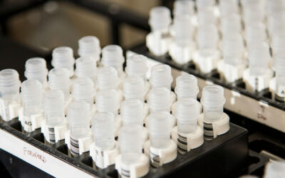
This published unit in Current Protocols in Stem Cell Biology illustrates a set of 7 protocols for culturing and dual labeling of human mesenchymal stem cells, preparing the cells for cellular iron and gadolinium quantification with inductively coupled mass spectrometry, detection of live and dead dual-labeled cells in agarose phantoms, and finally, a protocol for transplanting and imaging of these dual-labeled cells in vivo. Enabling timely and effective monitoring of stem cell delivery, migration and survival, these cutting-edge protocols are an advancement over the widely used single labeling with SPIONs that cannot not detect cell death.
Ultimately, this technique could facilitate efficient management and personalization of therapeutic cell-based regimens.
To find out more please read: Ngen, E. J., Kato, Y., & Artemov, D. (2017). Direct cell labeling to image transplanted stem cells in real time using a dual-contrast MRI technique. Current Protocols in Stem Cell Biology, 42, 5A.10.1–5A.10.19. doi: 10.1002/cpsc.33.