The inherent potential of stem cells to engender any cellular form and function drives the field of regenerative medicine. In recent years, stem cell research has witnessed prolific growth particularly in disease-modeling, drug discovery and patient-specific cell therapy. A major challenge in stem cell-mediated therapy is to understand and promote integration of transplanted stem cells into the host tissue. Fundamental to success of such therapies is development of robust methods to engraft stem cells and monitor their integration into the host network.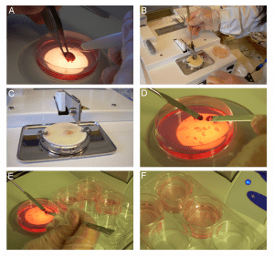
Central to this theme, David Forsberg, Eric Herlenius and co-workers at Karolinska Institute (Stockholm) and Mahidol University (Bangkok) have recently published a comprehensive set of cutting-edge protocols in Unit 2D.13 of Current Protocols in Stem Cell Biology. The unit outlines methods to culture slices of striatal and brainstem tissue (organotypic culture) for engraftment and integration of stem cells into the host neural network.
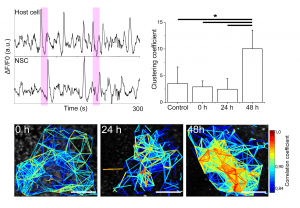
Integration of grafted neural stem cells into the host neural network
Organotypic cultures are 3-D model systems that can be used to investigate developmental and disease-related processes including morphological and functional cellular connections. The authors provide brief overview, effective illustrations and step-by-step guide to (i) establish organotypic cultures of brain by interface and rolling drum methods, (ii) prepare and engraft neural stem cells, (iii) introduce specific genes like cell-specific fluorescent markers by viral transfection, and (iv) measure structural and functional communication between engrafted stem cells and existing neural network by immunohistochemistry, time-lapse calcium imaging and direct injection of fluorescent gap-junction-permeable dye. The unit also includes details of necessary precautions, factors that increase the efficiency of engraftment, potential roadblocks, and critical parameters like pH, temperature, thickness of tissue slice, engrafted cell density and dye concentration that govern the overall success of the experiment.
This unit is a promising resource for establishing ex vivo 3-D tissue models for investigating temporal and spatial interactions between engrafted stem cells and host network. The unit is also an important element of the groundwork necessary for successful stem cell-mediated therapies in future.