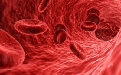
The development of bio-imaging techniques is of great interest to healthcare professionals. Techniques such as confocal microscopy, stimulated emission depletion microscopy, and stochastic optical reconstruction microscopy can be used for 3-dimensional imaging and for reconstruction of feature sizes below the incident light wavelength. However, these techniques require molecules to be marked with fluorescent labels in order to be imaged, and this can not only damage live cells but also influence cell physiology by mechanotransduction. A more suitable method for bio-imaging may be surface plasmon resonsance microscopy (SPRM) which does not require fluorescent labelling. It does, however, require optical couplers (prisms and gratings) and is intrisincally limited to studying very small regions of the order of several hundred nanometers, which is considerably below the size of a biological cell and therefore makes intracellular imaging impossible.

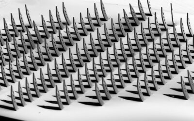
In Advanced Optical Materials this week, researchers from National Tsing Hua and National Yang-Ming Universities in Taiwan report a breakthrough SPRM bio-imagine device which is label-free, scalable and allows intracellular imaging. At the heart of the device is an array of split-ring resonators that have already been demostrated as refractive-index sensors. Exploiting this behaviour, the team has successfully demonstrated intracelluar imaging of human bone marrow-derived mesenchymal stem cells. Indeed this is the first reported device for intracellular plasmonic imaging by exciting multi-mode resonances in split-ring resonators.