 A device capable of simultaneous stimulation and recording of neural activity in the spinal cord may facilitate the restoration of motor and sensory functions in paralyzed patients following spinal cord injury. However, the biological complexity and soft, fragile structure of the spinal cord pose engineering challenges to develop such devices.
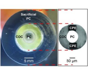
To address these design constraints, an MIT research group has developed highly flexible fiber probes, comprised entirely of polymers, for simultaneous optical stimulation and recording of neuronal activity. The fiber probes integrate optically-transparent polycarbonate and cyclic olefin copolymer that constitute the waveguide core and cladding, respectively, as well as conductive polyethylene electrodes.
The team report that these fiber probes exhibit low-loss light transmission even under repeated extreme bending deformation and enable optical stimulation and electrical recording of neural activity in the spinal cord of transgenic mice expressing a light sensitive protein channelrhodopsin 2. Furthermore, they showed that evoked spinal cord activity using the polymer fiber probes manifested in on-demand limb movements.